. 2005 Dec 15;438(7070):975-80.
doi: 10.1038/nature04293.

Probing ion-channel pores one proton at a time

Affiliations

Probing ion-channel pores one proton at a time

Gisela D Cymes et al. Nature. .

Abstract

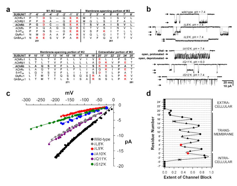
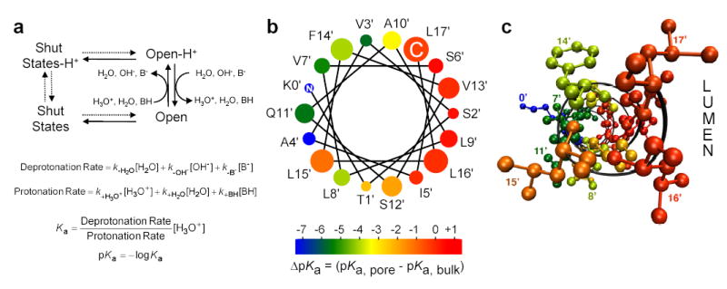
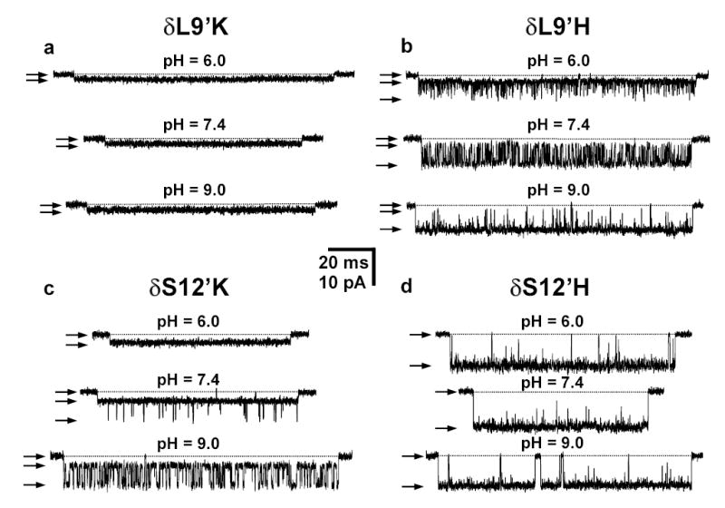
Although membrane proteins often rely on ionizable residues for structure and function, their ionization states under physiological conditions largely elude experimental estimation. To gain insight into the effect of the local microenvironment on the proton affinity of ionizable residues, we have engineered individual lysines, histidines and arginines along the alpha-helical lining of the transmembrane pore of the nicotinic acetylcholine receptor. We can detect individual proton binding-unbinding reactions electrophysiologically at the level of a single proton on a single side chain as brief blocking-unblocking events of the passing cation current. Kinetic analysis of these fluctuations yields the position-dependent rates of proton transfer, from which the corresponding pK(a) values and shifts in pK(a) can be calculated. Here we present a self-consistent, residue-by-residue description of the microenvironment around the pore-lining transmembrane alpha-helices (M2) in the open-channel conformation, in terms of the excess free energy that is required to keep the engineered basic side chains protonated relative to bulk water. A comparison with closed-channel data leads us to propose that the rotation of M2, which is frequently invoked as a hallmark of the gating mechanism of Cys-loop receptors, is minimal, if any.


Figures

Figure 1
Figure 1. Protonation of lysines causes partial channel block
a, Sequence alignment corresponding to the portion of M2 studied here. The four adult mouse-muscle AChR subunits (α1, β1, δ, and ɛ), and one representative each of the serotonin (5HT3), GABA, and glycine (GlyR) receptor-channel subunits are compared. Protonatable residues are indicated in red. AChR δ-subunit residues are indicated in bold. The prime-numbering system is indicated at the top, and the particular mouse δ-subunit residue numbers are at the bottom. b, Example single-channel inward currents (V ≅ −200 mV, [ACh] = 1 μM) recorded from HEK-293 cells expressing the indicated constructs. Openings are downward deflections. The shut (i.e., zero current) and the two open current-levels are indicated by arrows. The shut level of each trace is also indicated by a horizontal dotted line. Display fc = 6 kHz. c, Current-voltage (I–V) relationships for the five mutants in (b) and the wild-type AChR. For clarity, only the I–V curve corresponding to the blocked open state is shown for each mutant. d, The extent of channel block for each lysine construct was calculated as the conductance difference between the main level and the sublevel, normalized by the conductance of the main level (i.e., ΔConductance/Main-Level Conductance). For positions 0’ and 4’, however, the extent-of-block values (red squares) are predictions based on the values observed at neighboring positions (see text and Supplementary Methods for details). The solid line is a cubic-spline interpolation. Proposed membrane boundaries are tentative, and our data would not be inconsistent with these being displaced by approximately one turn toward the intracellular. Note that even residues that would be on the ‘back’ of the α-helix (e.g., 8’, 11’, 15’) exert a considerable electrostatic effect on the cation current. For most positions, the horizontal error bars (standard errors) are smaller than the experimental points. The data suggest that the pore’s lumen is to the right of the plot.
Figure 2
Figure 2. Experimental estimates of pKa-values
a, Conceptual framework of this work: a simple thermodynamic cycle. The channel interconverts among closed, desensitized (both referred here, collectively, as ‘shut states’), and open conformations with or without an extra proton bound to the pore domain. The association and dissociation of a single proton to the open state (but not to the shut states) is manifest as a discrete change in the rate of ion flow. Kinetic analysis of these fluctuations, and of the open ⇌ shut transitions, yields the rates of protonation and deprotonation of the open channel; these, along with the solution’s pH, are then combined to calculate the engineered-residue’s pKa. The three proton donors and three proton acceptors present in the solutions are indicated. BH and B- denote the protonated and deprotonated forms of the H+-buffer, respectively. Note that the kinetics of both proton transfer and channel shutting affect the duration of sojourns in the sub- and main levels (solid arrows). b, ΔpKa-values mapped onto an ideal α-helical wheel representation of δM2. The size of the symbols increases toward the extracellular end. Wild-type residues, and the C and N ends are indicated. The pore’s lumen would be to the right of the plot. The unique properties of the 15’ position are discussed in the Supplementary Discussion. c, ΔpKa-values mapped onto a ball-and-stick representation of the 0’ – 17’ δM2 stretch, using the atomic coordinates in the 1OED PDB file. The color code is the same as in (b). The backbone is indicated in ribbon representation. The molecular image was made with VMD.
Figure 3
Figure 3. pH-dependence of proton-transfer reactions
a, b, Example single-channel inward currents (V ≅ −100 mV, [ACh] = 1 μM) recorded from Leu-to-Lys (a) and Leu-to-His (b) δ9’ mutants at three different pH-values. The microenvironment around δ9’ slightly stabilizes the positive ɛNH3+ charge relative to bulk, and thus a lysine remains protonated in the 6.0 – 9.0 pH range. Hence, a histidine, with a lower proton affinity, is needed to reveal this position’s ΔpKa. As expected, the deprotonated state of the histidine is favored as the pH increases. c, d, Example single-channel inward currents (V ≅ −100 mV, [ACh] = 1 μM) recorded from Ser-to-Lys (c) and Ser-to-His (d) δ12’ mutants at three different pH-values. The microenvironment around δ12’ destabilizes the positive charge relative to bulk, and thus a histidine remains deprotonated in the 6.0 – 9.0 pH range. An arginine at this position, in contrast, still retains the labile proton, even at pH = 9.0 (data not shown). Thus, only a lysine, with an intermediate proton affinity, can reveal the ΔpKa at δ12’. As expected, the deprotonated state of the lysine is favored as the pH increases. Openings are downwards. The shut and the two open current-levels are indicated by arrows. The shut level is also indicated by a horizontal dotted line. Display fc = 6 kHz.

References

    1. Perutz MF. Electrostatic effects in proteins. Science. 1978;201:1187–1191. - PubMed
    1. Warshel A. Electrostatic basis of structure-function correlation in proteins. Acc Chem Res. 1981;14:284–290.
    1. Davis ME, McCammon JA. Electrostatics in biomolecular structure and dynamics. Chem Rev. 1990;90:509–521.
    1. Honig B, Nicholls A. Classical electrostatics in biology and chemistry. Science. 1995;268:1144–1149. - PubMed
    1. Imoto K, et al. Rings of negatively charged amino acids determine the acetylcholine receptor channel conductance. Nature. 1988;335:645–648. - PubMed

Publication types

MeSH terms