

Effects of sex and age on bone microstructure at the ultradistal radius: a population-based noninvasive in vivo assessment
- PMID: 16355281
- PMCID: PMC1352156
- DOI: 10.1359/JBMR.050916
Effects of sex and age on bone microstructure at the ultradistal radius: a population-based noninvasive in vivo assessment
Abstract
In a population-based cross-sectional study, we examined effects of sex and age on bone microstructure at the wrist using high-resolution 3-D pQCT. Compared with women, men had thicker trabeculae in young adulthood and had less microstructural damage with aging. These findings may contribute to the virtual immunity of men to age-related increases in wrist fractures.
Introduction: Although changes in bone microstructure contribute to fracture risk independently of BMD, it has not heretofore been possible to assess this noninvasively in population-based studies.
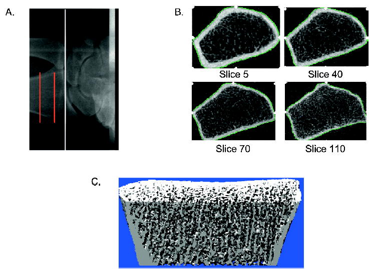
Materials and methods: We used high-resolution 3-D pQCT imaging (voxel size, 89 mum) to define, in a random sample of women (n = 324) and men (n = 278) 21-97 years of age, sex and age effects on bone microstructure at the wrist.
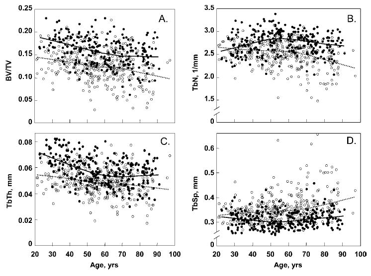
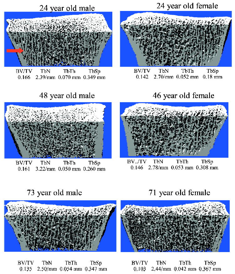
Results: Relative to young women (age, 20-29 years), young men had greater trabecular bone volume/tissue volume (BV/TV; by 26%, p = 0.001) and trabecular thickness (TbTh; by 28%, p < 0.001) but similar values for trabecular number (TbN) and trabecular separation (TbSp). Between ages 20 and 90 years, cross-sectional decreases in BV/TV were similar in women (-27%) and in men (-26%), but whereas women had significant decreases in TbN (-13%) and increases in TbSp (+24%), these parameters had little net change over life in men (+7% and -2% for TbN and TbSp, respectively; p < 0.001 versus women). However, TbTh decreased to a greater extent in men (-24%) than in women (-18%; p = 0.010 versus men).
Conclusions: Whereas decreases with age in trabecular BV/TV are similar in men and women, the structural basis for the decrease in trabecular volume is quite different between the sexes. Thus, over life, women undergo loss of trabeculae with an increase in TbSp, whereas men begin young adult life with thicker trabeculae and primarily sustain trabecular thinning with no net change in TbN or TbSp. Because decreases in TbN have been shown to have a much greater impact on bone strength compared with decreases in TbTh, these findings may help explain the lower life-long risk of fractures in men, and specifically, their virtual immunity to age-related increases in distal forearm fractures.
Conflict of interest statement
Dr Melton has received speaker’s honoraria from Procter & Gamble, Merck, and Amgen. All other authors have no conflict of interest.
Figures
References
-
- Miller PD, Zapalowski C, Kulak CAM, Bilezikian JP. Bone densitometry: The best way to detect osteoporosis and to monitor therapy. J Clin Endocrinol Metab. 1999;84:1867–1871. - PubMed
-
- Riggs BL, Melton LJI, Robb RA, Camp JJ, Atkinson EJ, Peterson JM, Rouleau PA, McCollough CH, Bouxsein ML, Khosla S. Population-based study of age and sex differences in bone volumetric density, size, geometry, and structure at different skeletal sites. J Bone Miner Res. 2004;19:1945–1954. - PubMed
-
- Bouxsein ML 2001 Biomechanics of age-related fractures. In: Marens R, Kelsey J, Feldman D (eds). Osteoporosis, 2nd ed. Academic Press, San Diego, CA, USA, pp. 509–534.
-
- Aaron JE, Makins NB, Sagreiya K. The microanatomy of trabecular bone loss in normal aging men and women. Clin Orthop. 1987;215:260–271. - PubMed
Publication types
MeSH terms
Grants and funding
LinkOut - more resources
Full Text Sources
Other Literature Sources
Medical
