Review
. 2005 Nov-Dec;7(11-12):1568-80.
doi: 10.1089/ars.2005.7.1568.

Role of poly(ADP-ribose) polymerase-1 activation in the pathogenesis of diabetic complications: endothelial dysfunction, as a common underlying theme

Affiliations
Review

Role of poly(ADP-ribose) polymerase-1 activation in the pathogenesis of diabetic complications: endothelial dysfunction, as a common underlying theme

Pál Pacher et al. Antioxid Redox Signal. 2005 Nov-Dec.

Abstract

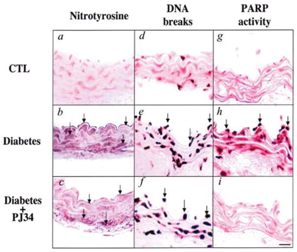
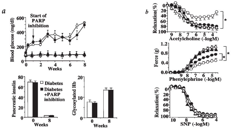
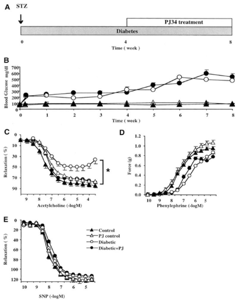
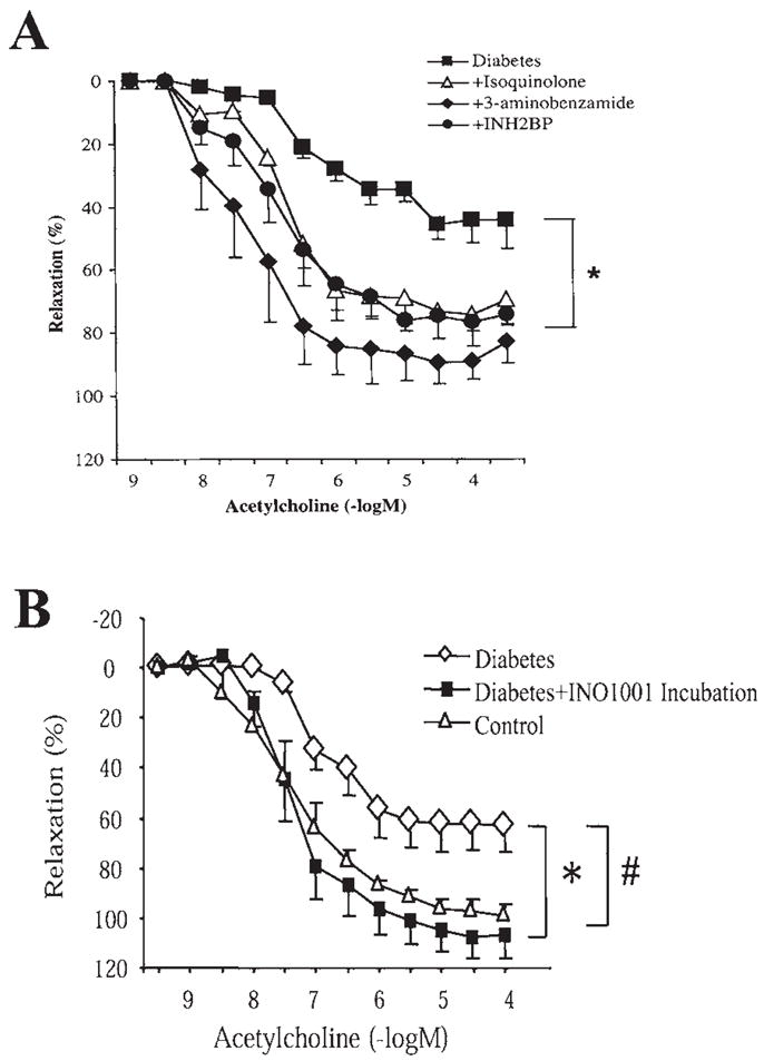
Hyperglycemia-induced overproduction of superoxide by mitochondrial electron-transport chain triggers several pathways of injury involved in the pathogenesis of diabetic complications [protein kinase C (PKC), hexosamine and polyol pathway fluxes, advanced glycation end product (AGE) formation] by inhibiting glyceraldehyde- 3-phosphate dehydrogenase (GAPDH) activity. Increased oxidative and nitrosative stress activates the nuclear enzyme, poly(ADP-ribose) polymerase-1 (PARP). PARP activation, on the one hand, depletes its substrate, NAD+, slowing the rate of glycolysis, electron transport, and ATP formation. On the other hand, it inhibits GAPDH by poly(ADP-ribosy)lation. These processes result in acute endothelial dysfunction in diabetic blood vessels, which importantly contributes to the development of various diabetic complications. Accordingly, hyperglycemia-induced activation of PKC isoforms, hexosaminase pathway flux, and AGE formation is prevented by blocking PARP activity. Furthermore, inhibition of PARP protects against diabetic cardiovascular dysfunction in preclinical models. PARP activation is present in microvasculature of human diabetic subjects. The oxidative/nitrosative stress-PARP pathway leads to diabetes-induced endothelial dysfunction, which may be an important underlying mechanism for the pathogenesis of other diabetic complications (cardiomyopathy, nephropathy, neuropathy, and retinopathy). This review focuses on the role of PARP in diabetic complications and the unique therapeutic potential of PARP inhibition in the prevention or reversal of diabetic complications.


Figures

FIG. 1
FIG. 1. Reactive nitrogen species generation, DNA breakage, and PARP activation in diabetic blood vessels
(a–c) Immunohistochemical staining for nitrotyrosine in control rings (a), in rings from diabetic mice treated with vehicle at 8 weeks (b), and in rings from diabetic mice treated with PJ34 (c). (d–f) Terminal deoxyribonucleotidyl transferase-mediated dUTP nick-end labeling, an indicator of DNA-strand breakage, in control rings (d), in rings from diabetic mice treated with vehicle at 8 weeks (e), and in rings from diabetic mice treated with PJ34 (f). (g–i) Immunohistochemical staining for poly(ADP-ribose), an indicator of PARP activation, in control rings (g), in rings from diabetic mice treated with vehicle at 8 weeks (h), and in rings from diabetic mice treated with PJ34 (i). Reproduced with permission from 33.
FIG. 2
FIG. 2. Reversal of diabetes-induced endothelial dysfunction by pharmacological inhibition of PARP
The following symbols were used for the respective groups: animals that received no STZ injection (Δ), nondiabetic control animals at 8 weeks treated with PJ34 between week 1 and 8 (▴), diabetic animals at 8 weeks treated with vehicle (▲), diabetic animals at 8 weeks treated with PJ34 between week 1 and 8 (●). (a) Blood glucose levels, pancreatic insulin content (ng of insulin/mg of pancreatic protein), and blood glycosylated hemoglobin (Hb) (expressed as % of total Hb) at 0–8 weeks in nondiabetic, control male BALB/c mice, and at 0–8 weeks after STZ treatment (diabetic) in male BALB/c mice. PARP inhibitor treatment, starting at 1 week after STZ and continuing until the end of week 8, is indicated by the arrow. Pancreatic insulin and glycated hemoglobin levels are shown at 8 weeks in vehicle-treated and STZ-treated animals, in the presence or absence of PJ34 treatment. (b) Acetylcholine-induced, endothelium-dependent relaxations, phenylephrine-induced contractions, and sodium nitroprusside (SNP)-induced endothelium-independent relaxations. *p < 0.05 for vehicle-treated diabetic versus PJ34-treated diabetic mice (n = 8 per group). Reproduced with permission from 33.
FIG. 3
FIG. 3. Pharmacological inhibition of PARP restores impaired endothelium-dependent relaxant ability of the diabetic vessels
Blood glucose levels and vascular responsiveness are presented. Endothelium-dependent relaxations were induced by acetylcholine, contractions induced by phenylephrine, and endothelium-independent relaxations induced by sodium nitroprusside (SNP) in control (nondiabetic) male Balb/c mice and 1, 4, and 8 weeks after STZ-induced diabetes. Vehicle or PARP inhibitor (PJ34, 10 mg/kg oral gavage once a day) treatment started at 4 weeks after STZ and continued until 8 weeks (the end of the experimental period). There was a marked and selective impairment of the endothelium-dependent relaxant ability of the vascular rings in diabetes at 4 and 8 weeks. Treatment with the PARP inhibitor between weeks 4 and 8 restored to normal the endothelium-dependent relaxant ability of the diabetic vessels despite the persistence of hyperglycemia. *p < 0.05 for differences between experimental groups, as indicated. n = 8 per group. Reproduced with permission from 91.
FIG. 4
FIG. 4. In vitro treatment with all PARP inhibitors improved the endothelium-dependent relaxant ability of the diabetic vessels
(A) Endothelium-dependent relaxations induced by acetylcholine in control (nondiabetic) male Balb/c mice and 4 weeks after STZ-induced diabetes. In a subgroup of the vascular rings, evaluation of vascular responsiveness was preceded by 1-h incubation with three structurally different PARP inhibitors: 3-aminobenzamide (3 mmol/L), 5-iodo-6-amino-1,2-benzopyrone (INH2BP) (100 μmol/L), or 1,5-dihydroxyisoquinoline (Isoquinolone) (30 μmol/L). There was a marked and selective impairment of the endothelium-dependent relaxant ability of the vascular rings in diabetes at 4 weeks. In vitro treatment with all PARP inhibitors improved the endothelium-dependent relaxant ability of the diabetic vessels. *p < 0.05 for differences between experimental groups, as indicated. n = 8 per group. Reproduced with permission from 91. (B) Endothelium-dependent relaxations induced by acetylcholine in control (nondiabetic) male Balb/c mice and 6 weeks after STZ-induced diabetes. In a subgroup of the vascular rings, evaluation of vascular responsiveness was preceded by 1-h incubation with the novel potent PARP inhibitor, INO1001 (3 μmol/L). There was a marked and selective impairment of the endothelium-dependent relaxant ability of the vascular rings in diabetes at 6 weeks. In vitro treatment with all PARP inhibitors improved the endothelium-dependent relaxant ability of the diabetic vessels. #, *p < 0.05 for differences between experimental groups, as indicated. n = 8 per group.
FIG. 5
FIG. 5. Reversal of diabetes-induced endothelial dysfunction by pharmacological inhibition of PARP in diabetic NOD mouse vascular rings
Epinephrine-induced contractions (upper panel), acetylcholine-induced endothelium-dependent relaxation (middle panel), and sodium nitroprusside (SNP)-induced endothelium-independent relaxations (lower panel).■, control; ○, control + PJ34; □, diabetes; ●, diabetes + PJ34. Each point of the curve represents the mean ± SE of five to eight experiments in vascular rings. *p < 0.05 versus control; #p < 0.05 versus diabetes. Reproduced with permission from 72.
FIG. 6
FIG. 6. Overview of the role of PARP in regulating multiple components of hyperglycemia-induced endothelial dysfunction
High circulating glucose interacts with the vascular endothelium where it triggers the release of oxidant mediators from the mitochondrial electron transport chain, as well as from NADH/NADPH oxidase and other sources. NO, in turn, combines with superoxide (O2 to yield peroxynitrite (ONOO). Hydroxyl radical (OH·) (produced from superoxide via the iron-catalyzed Haber–Weiss reaction) and peroxynitrite or peroxynitrous acid induce the development of DNA single-strand breakage, with consequent activation of PARP. Depletion of the cellular NAD+ leads to inhibition of cellular ATP-generating pathways leading to cellular dysfunction. The PARP-triggered depletion of cellular NADPH directly impairs the endothelium-dependent relaxations. The effects of elevated glucose are also exacerbated by increased aldose reductase activity leading to depletion of NADPH and generation of reactive oxidants. NO alone does not induce DNA single-strand breakage, but may combine with superoxide (produced from the mitochondrial chain or from other cellular sources) to yield peroxynitrite. Under conditions of low cellular L-arginine, NOS may produce both superoxide and NO, which then can combine to form peroxynitrite. PARP activation, via a not yet characterized fashion, can promote the activation of nuclear factor-κB, AP-1, mitogen-activated protein (MAP) kinases, and the expression of proinflammatory mediators, adhesion molecules, and iNOS. PARP activation contributes to the activation of PKC. PARP activation also leads to the inhibition of cellular GAPDH activity, at least in part via the direct poly(ADP-ribosyl)ation of GAPDH. PARP-independent, parallel pathways of cellular metabolic inhibition can be activated by NO, hydroxyl radical, superoxide, and peroxynitrite.

References

    1. Affar EB, Shah RG, Dallaire AK, Castonguay V, Shah GM. Role of poly(ADP-ribose) polymerase in rapid intracellular acidification induced by alkylating DNA damage. Proc Natl Acad Sci U S A. 2002;99:245–250. - PMC - PubMed
    1. Anderson D, Yu TW, Wright J, Ioannides C. An examination of DNA strand breakage in the comet assay and antioxidant capacity in diabetic patients. Mutat Res. 1998;398:151–161. - PubMed
    1. Andreoli SP. Mechanisms of endothelial cell ATP depletion after oxidant injury. Pediatr Res. 1989;25:97–101. - PubMed
    1. Astley S, Langrish-Smith A, Southon S, Sampson M. Vitamin E supplementation and oxidative damage to DNA and plasma LDL in type 1 diabetes. Diabetes Care. 1999;22:1626–1631. - PubMed
    1. Beckman JA. Inhibition of protein kinase Cbeta prevents impaired endothelium-dependent vasodilation caused by hyperglycemia in humans. Circ Res. 2002;90:107–111. - PubMed

MeSH terms

Substances