Review
. 2005;9(6):607-21.
doi: 10.1186/cc3877. Epub 2005 Oct 18.

Clinical review: Positive end-expiratory pressure and cardiac output

Affiliations
Review

Clinical review: Positive end-expiratory pressure and cardiac output

Thomas Luecke et al. Crit Care. 2005.

Abstract

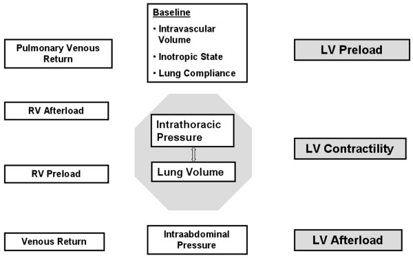
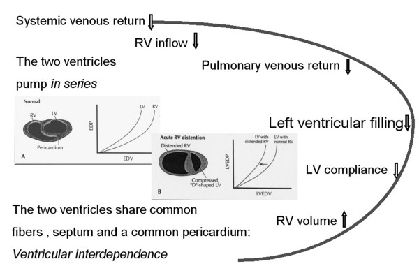
In patients with acute lung injury, high levels of positive end-expiratory pressure (PEEP) may be necessary to maintain or restore oxygenation, despite the fact that 'aggressive' mechanical ventilation can markedly affect cardiac function in a complex and often unpredictable fashion. As heart rate usually does not change with PEEP, the entire fall in cardiac output is a consequence of a reduction in left ventricular stroke volume (SV). PEEP-induced changes in cardiac output are analyzed, therefore, in terms of changes in SV and its determinants (preload, afterload, contractility and ventricular compliance). Mechanical ventilation with PEEP, like any other active or passive ventilatory maneuver, primarily affects cardiac function by changing lung volume and intrathoracic pressure. In order to describe the direct cardiocirculatory consequences of respiratory failure necessitating mechanical ventilation and PEEP, this review will focus on the effects of changes in lung volume, factors controlling venous return, the diastolic interactions between the ventricles and the effects of intrathoracic pressure on cardiac function, specifically left ventricular function. Finally, the hemodynamic consequences of PEEP in patients with heart failure, chronic obstructive pulmonary disease and acute respiratory distress syndrome are discussed.


Figures

Figure 1
Figure 1
Schematic representation of potential cardiopulmonary interactions with changes in intrathoracic pressure (ITP) and lung volume (redrawn with permission from [137]). To obtain a more focused view of these numerous interactions, one can simply group all hemodynamic effects of ventilation into processes that, by changing lung volume and ITP, affect left ventricular (LV) preload, contractility and afterload [6]. RV, right ventricular.
Figure 2
Figure 2
Effects of positive end-expiratory pressure (PEEP) on venous return and cardiac output. (a) Theoretical effects of PEEP on venous return (VR) and cardiac output (CO). PEEP causes an increase in intrathoracic pressure (ITP) and a right shift in the cardiac function curve. If there were no change in the VR curve, then CO and VR would decrease (from point A to point B). However, if there is a compensatory increase in mean systemic pressure (from Pms1 to Pms2), then the system will exist in equilibrium at point C, at which VR and CO would be maintained compared to zero end-expiratory pressure (ZEEP) conditions. Pms can increase either by an increase in stressed volume or sympathoadrenal stimulation. (b) Another possible scheme for the changes in VR with PEEP. If there is an increase in the pressure at which flow limitation occurs, then the ability of an increase in Pms to buffer PEEP-induced decreases in VR is markedly less. FL1, flow limiting point at ZEEP; FL2, flow limiting point at PEEP. Modified from [8], with permission.
Figure 3
Figure 3
Characteristic echocardiographic patterns of acute cor pulmonale with transesophageal echocardiography. In the upper panel, right ventricular (RV) dilation is observed in a long-axis view, at end-diastole (left) and end-systole (right). Also note the reduced size of the left ventricle (LV). In the lower panel, septal dyskinesia is observed, in the same patient, in a short-axis view: at end-systole/early diastole (right) the interventricular septum (IVS) is shifted toward the LV cavity center, and the septal curvature is inverted (arrow). TP, tracheal pressure. Reproduced from [63], with permission.
Figure 4
Figure 4
Effect of positive end-expiratory pressure (PEEP) on left ventricular (LV) filling. Any decrease in systemic venous return and, thus, right ventricular (RV) inflow must result in decreased pulmonary venous return and inflow to the left ventricle because the two ventricles pump in series. In addition to this passive coupling of the right and left ventricle, PEEP may have more direct mechanical effects on LV filling as the two ventricles share common fiber bundles, a common interventricular septum, coexist within the same pericardial space, and are surrounded by a fixed cardiac fossa volume. This parallel interaction between the ventricles, whereby the function of one ventricle influences the function of the other, is called ventricular interdependence. Classically, ventricular interdependence is thought to occur as increases in RV volume decrease LV diastolic compliance, LV preload, and LV output (acute cor pulmonale). EDP, end-diastolic pressure; EDV end-diastolic volume. Inserts adapted from [69], with permission.
Figure 5
Figure 5
Effects of continuous positive-pressure ventilation on the end-diastolic (ED) and end-systolic (ES) volume (V)-transmural pressure (tm) relationship of the left ventricle (LV). Closed circles represent the mean V-tm coordinates at low levels of positive end-expiratory pressure (PEEP; 0, 5, 10 cmH2O), and the continuous lines are drawn through these points to indicate typical V-tm curves. Open circles represent the mean V-tm coordinates at high levels of PEEP (15, 20, and 20 cmH2O) with volume expansion. Both ESV and EDP are reduced at the same pressures, indicating a leftward displacement of the V-tm pressure curves at high levels of PEEP. Reproduced from [83], with permission.
Figure 6
Figure 6
Starling relationship between cardiac output (CO) and the end-diastolic volume (EDV) of the right ventricle (RV; right curve) and left ventricle (LV; left curve) as airway pressure was progressively increased from 0 (upper right data point) to 20 cmH2O (lower left data point). Note that volume expansion at a positive end-expiratory pressure (PEEP) of 20 cmH2O (20 + VE) entirely reversed the decrease in RV EDV and LV EDV and restored CO. VE, volume expansion. Reproduced from [83], with permission.

Similar articles

Cited by

References

    1. Dreyfuss D, Saumon G. Ventilator-induced lung injury. Lessons from experimental studies. Am J Respir Crit Care Med. 1998;157:294–330. - PubMed
    1. Maggiore SM, Jonson B, Richard JC, Jaber S, Lemaire F, Brochard L. Alveolar derecruitment at decremental positive end-expiratory pressure levels in acute lung injury: comparison with the lower inflection point, oxygenation, and compliance. Am J Respir Crit Care Med. 2001;164:795–801. - PubMed
    1. Rouby JJ, Lu Q, Goldstein I. Selecting the right level of positive end-expiratory pressure in patients with acute respiratory distress syndrome. Am J Respir Crit Care Med. 2002;165:1182–1186. - PubMed
    1. Cournand A, Motley HL, Werko L, Richards D. Physiological studies of the effects of intermittent positive pressure breathing on cardiac output in man. Am J Physiol. 1948;152:162–174. - PubMed
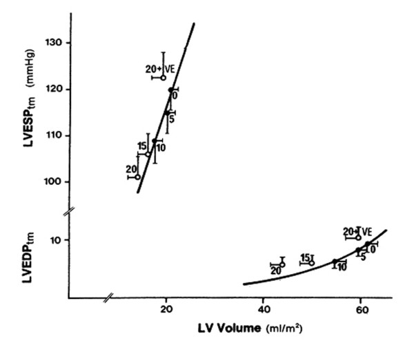
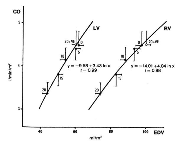
    1. Viquerat CE, Righetti A, Suter PM. Biventricular volumes and function in patients with adult respiratory distress syndrome ventilated with PEEP. Chest. 1983;83:509–514. - PubMed