Comparative Study
. 2006 Jan;5(1):17-25.
doi: 10.1016/j.jcf.2005.11.001. Epub 2005 Dec 13.

Attenuation of host defense function of lung phagocytes in young cystic fibrosis patients

Affiliations
Comparative Study

Attenuation of host defense function of lung phagocytes in young cystic fibrosis patients

Neil E Alexis et al. J Cyst Fibros. 2006 Jan.

Abstract

Background: Recent reports suggest that endotoxin exposure can blunt phagocyte functions. The aim of this study was to examine whether lung phagocytic cells have altered host defense function in young cystic fibrosis (CF) patients, and to explore the contribution of neutrophil elastase (NE) and surfactant proteins to these effects.

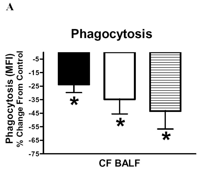
Methods: BALF cells from CF children (N=12) and disease controls (N=12) were analyzed by flow cytometry for mCD14 and HLA-DR expression and phagocytosis. The effects of exogenous surfactant protein A and D (SP-A,D) and proteases on BALF cells in short term culture were assessed experimentally.

Results: Expression of the surface markers mCD14 and HLA-DR, and phagocytosis, were all blunted on CF phagocytes compared to disease controls (p<0.05). In CF phagocytes, SP-A enhanced both phagocytosis and mCD14 expression (p<0.05). Both CF BALF and NE reduced phagocytosis and expression of mCD14 and HLA-DR (p<0.05) by non-CF phagocytes; the latter effect was attenuated by protease inhibitor.

Conclusion: CF airway phagocytes appear to have altered host defense functions that could contribute to poor bacterial clearance. These impairments can be reproduced by incubation of non-CF cells with NE, while SP-A can partially reverse them. Decreasing protease activity and increasing collectin activity may be beneficial in early CF.


Figures

Figure 1
Figure 1
mCD14 (A) and HLA-DR (B) expression (MFI) on CF (N=12, solid bar) and non-CF (N=12, open bar) BALF macrophages and monocytes. mCD14 and HLA-DR expression were significantly (*p<0.05) decreased in CF versus non-CF subjects.
Figure 2
Figure 2
Phagocytosis of zymosan particles by macrophages and neutrophils recovered from BALF of CF (N=11, solid bar) and non-CF (N=6, open bar) subjects as measured by both mean fluorescence intensity (MFI) (2A) and % cells (macrophages, neutrophils) that took up zymosan particles (2B). Phagocytosis on macrophages and neutrophils is significantly (* p<0.05) decreased in the CF (solid bar) vs. non-CF (open bar) subject groups. Figures 2C and 2D show representative histograms of a non-CF and CF subject, respectively. Macrophage phagocytosis after zymosan exposure is shown as the dark histogram. No zymosan exposure is shown as the grey histogram. The M2 region indicates zymosan uptake by macrophages and M1 (control region) indicates no zymosan uptake.
Figure 2
Figure 2
Phagocytosis of zymosan particles by macrophages and neutrophils recovered from BALF of CF (N=11, solid bar) and non-CF (N=6, open bar) subjects as measured by both mean fluorescence intensity (MFI) (2A) and % cells (macrophages, neutrophils) that took up zymosan particles (2B). Phagocytosis on macrophages and neutrophils is significantly (* p<0.05) decreased in the CF (solid bar) vs. non-CF (open bar) subject groups. Figures 2C and 2D show representative histograms of a non-CF and CF subject, respectively. Macrophage phagocytosis after zymosan exposure is shown as the dark histogram. No zymosan exposure is shown as the grey histogram. The M2 region indicates zymosan uptake by macrophages and M1 (control region) indicates no zymosan uptake.
Figure 3
Figure 3
BALF cell culture experiments measuring phagocytosis (3A) and mCD14 expression (3B) on macrophages obtained from 12 CF subjects 24h after incubation with SP-A, SP-D, or LPS. Phagocytosis (MFI, % change from control) was significantly increased following SP-A and SP-D incubation (*p<0.05). mCD14 expression (MFI, % change from control) was significantly increased following incubation with SP-A (*p<0.05).
Figure 4
Figure 4
BALF cell culture experiments measuring phagocytosis (4A), mCD14 (4B) and HLA-DR (4C) expression on non-CF BALF cells after in vitro incubations with cell free CF BALF, neutrophil elastase (NE) and these conditions with addition of proteinase inhibitor (PI). 4A: Phagocytosis was measured in macrophages (solid bar), monocytes (open bar) and neutrophils (horizontal hatch). Phagocytosis (MFI, % change from control) was significantly (*p<0.05) reduced for macrophages, monocytes and neutrophils following incubation with CF BALF (N=5); other conditions NS and not shown. 4B and 4C show surface marker expression after BALF + PI (N=5), NE (N=4) and NE + PI (N=4). Both, mCD14 (4B) and HLA-DR (4C) expression (MFI, as % change from control) were significantly decreased following incubation with CF BALF and NE (*p<0.05). PI significantly (#p<0.05) attenuated the NE-induced decrement in mCD14 and HLA-DR expression, but not the CF BALF-induced decrement.
Figure 4
Figure 4
BALF cell culture experiments measuring phagocytosis (4A), mCD14 (4B) and HLA-DR (4C) expression on non-CF BALF cells after in vitro incubations with cell free CF BALF, neutrophil elastase (NE) and these conditions with addition of proteinase inhibitor (PI). 4A: Phagocytosis was measured in macrophages (solid bar), monocytes (open bar) and neutrophils (horizontal hatch). Phagocytosis (MFI, % change from control) was significantly (*p<0.05) reduced for macrophages, monocytes and neutrophils following incubation with CF BALF (N=5); other conditions NS and not shown. 4B and 4C show surface marker expression after BALF + PI (N=5), NE (N=4) and NE + PI (N=4). Both, mCD14 (4B) and HLA-DR (4C) expression (MFI, as % change from control) were significantly decreased following incubation with CF BALF and NE (*p<0.05). PI significantly (#p<0.05) attenuated the NE-induced decrement in mCD14 and HLA-DR expression, but not the CF BALF-induced decrement.

Similar articles

Cited by

References

    1. Chmiel JF, Berger M, Konstan MW. The role of inflammation in the pathophysiology of CF lung disease. Clin Rev Allergy Immunol. 2002;23:5–27. - PubMed
    1. Bruce MC, Poncz L, Klinger JD, Stern RC, Tomashefski JF, Jr, Dearborn DG. Biochemical and pathologic evidence for proteolytic destruction of lung connective tissue in cystic fibrosis. Am Rev Respir Dis. 1985;132:529–535. - PubMed
    1. Cantin A. Cystic fibrosis lung inflammation: early, sustained, and severe. Am J Respir Crit Care Med. 1995;151(4):939–941. - PubMed
    1. Doring G, Worlitzsch D. Inflammation in cystic fibrosis and its management. Paediatr Respir Rev. 2000;1(2):101–106. - PubMed
    1. Berger M. Inflammatory mediators in cystic fibrosis lung disease. Allergy Asthma Proc. 2002;23(1):19–25. - PubMed

Publication types

MeSH terms