. 2005 Dec 14;33(22):7074-89.
doi: 10.1093/nar/gki1014. Print 2005.

The autoregulatory translational control element of poly(A)-binding protein mRNA forms a heteromeric ribonucleoprotein complex

Affiliations

The autoregulatory translational control element of poly(A)-binding protein mRNA forms a heteromeric ribonucleoprotein complex

Gopal P Patel et al. Nucleic Acids Res. .

Abstract

Repression of poly(A)-binding protein (PABP) mRNA translation involves the binding of PABP to the adenine-rich autoregulatory sequence (ARS) in the 5'-untranslated region of its own mRNA. In this report, we show that the ARS forms a complex in vitro with PABP, and two additional polypeptides of 63 and 105 kDa. The 63 and 105 kDa polypeptides were identified, as IMP1, an ortholog of chicken zip-code binding polypeptide, and UNR, a PABP binding polypeptide, respectively, by mass spectrometry of the ARS RNA affinity purified samples. Using a modified ribonucleoprotein (RNP) immunoprecipitation procedure we further show that indeed, both IMP1 and UNR bind to the ARS containing reporter RNA in vivo. Although both IMP1 and UNR could bind independently to the ARS RNA in vitro, their RNA-binding ability was stimulated by PABP. Mutational analyses of the ARS show that the presence of four of the six oligo(A) regions of the ARS was sufficient to repress translation and the length of the conserved pyrimidine spacers between the oligo(A) sequences was important for ARS function. The ability of mutant ARS RNAs to form the PABP, IMP1 and UNR containing RNP complex correlates well with the translational repressor activity of the ARS. There is also a direct relationship between the length of the poly(A) RNAs and their ability to form a trimeric complex with PABP, and to repress mRNA translation. UV crosslinking studies suggest that the ARS is less efficient than a poly(A) RNA of similar length, to bind to PABP. We show here that the ARS cannot efficiently form a trimeric complex with PABP; therefore, additional interactions with IMP1 and UNR to form a heteromeric RNP complex may be required for maximal repression of PABP mRNA translation under physiological conditions.


Figures
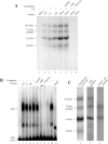
Figure 1
Figure 1
Formation of ARS RNA–protein complex. (A) RNA–protein crosslinking by UV. The in vitro synthesized [32P]-labeled ARS RNA (≈3 ng, 3 × 10⁵ c.p.m.) was incubated with different cell extract (≈60 µg protein) in the flat cap of a 0.2 ml PCR tube. Following the UV treatment, the samples were treated with RNaseA/RNase T1, analyzed by 10% SDS–PAGE and autoradiographed as described under experimental procedures. Cell extracts from mouse NIH3T3 fibroblasts (lane 2) and C2 myoblasts (lane 3), and human HEK293 (lane 4) and HeLa cells (lane 5) were used for these studies. One sample containing HeLa cell extract (lane 1) was analyzed without UV treatment as a control. Approximately 300 ng of the non-radioactive pGEM-T (lane 6), ARS (lane 7) or poly(A)50 RNA (lane 8) was used for competition studies. (B) Analysis of RNP complex by REMSA. The in vitro synthesized [32P]-labeled ARS RNA (≈1 ng, 1 × 10⁵ c.p.m.) was incubated with different cell extracts (20 µg) as indicated above each lane. The unbound RNA was digested with RNaseT1 and subjected to electrophoresis in a 2% agarose as described in the experimental procedures. Lane 1, radioactive ARS RNA incubated without the cell extract; lanes 2–5, radioactive ARS RNA incubated with NIH3T3, C2, HEK293 and HeLa cell extracts, respectively. Lanes 6–8, competition with 100 ng of non-radioactive pGEM-T, ARS, or poly(A)50 RNA, respectively. Lane 9, cell extract was pre-incubated with the PABP antibody and precleared with protein A-sepharose beads before being used for REMSA. Approximately 20 µg of PABP deficient cell extract was used for REMSA. Lane 10, cell extract was similarly treated with the GFP antibody (BD Biosciences) before being used for REMSA. Lane 11, radiolabeled ARS RNA (≈ 1 ng, 1 × 10⁵ c.p.m.) incubated with ≈2 ng of purified 6×His-PABP. (C) Analysis of RNP complexes by SDS–PAGE. RNP complex formation was initiated as described above and the samples were irradiated by UV before being resolved in a 2% agarose gel. The RNP bands (as shown in Figure 1B) were excised from the gel, treated with RNaseA/RNase T1, and analyzed by 10% SDS–PAGE as described in the Materials and Methods. Lane 1, ARS RNA and HeLa extracts treated with UV and analyzed before gel purification. Lane 2, polypeptides from the gel purified slower migrating ARC. Lane 3, polypeptides from the faster migrating minor complex.
Figure 2
Figure 2
Comparison of the ability of the ARS and poly(A) RNA to bind PABP. (A) Gel-shift assays of binding of PABP to the ARS and poly(A) RNAs of various length. REMSA was performed using 0.2, 0.4, 0.8 and 1.6 ng of purified 6× His-PABP and ≈1.5 ng of [32P]-labeled ARS (lanes 2–5), poly(A)50 (lanes 6–9), poly(A)20 (lanes 10–13) and poly(A)13 (lanes 14–17) RNAs. Samples were analyzed on 5% PAGE under non-denaturing conditions. (B and C) UV crosslinking assays of binding of PABP to ARS and poly(A)50 RNAs. The in vitro synthesized [32P]-labeled ARS and poly(A)50 RNAs (≈3 ng, 3 × 10⁵) were incubated with the purified PABP (≈2 ng) for 5 min at room temperature. Unlabeled ARS (lanes 3–8) or poly(A)50 (lanes 9–14) competitor RNAs were added (10-fold molar excess increment) and incubated further at the room temperature for 3 min. Following the UV treatment, the samples were treated with RNase A/RNase T1, fractionated on a 10% SDS–PAGE and visualized by autoradiography. Lane 1, samples without UV treatment; lane 2, samples without unlabeled competitor RNA. (D) The RNP bands in (A and B) were excised by superimposing the radiograph and the level of radioactivity was measured by scintillation counter. The average level of radioactivity of the RNP complex in each band from three separate competition experiments was plotted against the molar concentration of the competitor RNA.
Figure 3
Figure 3
Affinity chromatography of ARS RNA-binding proteins. In vitro synthesized ARS RNA was covalently linked to agarose beads and incubated with HeLa cell extract. The bound polypeptides were eluted, resolved on 10% SDS–PAGE and visualized by silver staining. Lane 1, pGEM-T and lane 2, ARS RNA bound protein fractions. The polypeptides specific for the ARS RNA affinity chromatography are shown by arrows. The polypeptides with bold arrows were seen in the UV crosslinking experiments.
Figure 4
Figure 4
Interaction of IMP1 and UNR with RNA. (A) Presence of IMP1, PABP and UNR in the ARS RNA–protein complex. REMSA was performed using [32P]-labeled ARS RNA and HeLa cell extract as described. Cell extract (≈20 µg) was incubated with ≈1 µg of either non-immunized serum (lane 3), IMP1 (lane 4), PABP (lane 5) or UNR (lane 6) antibody for 10 min on ice prior to the addition of the labeled ARS RNA (≈1 ng, 1 × 10⁵ c.p.m.). Samples were subjected to 5% PAGE under non-denaturing conditions. (B) Binding of IMP1 to the ARS RNA. [35S]methionine labeled 6× His-tagged IMP1, PABP, UNR and β-galactosidase were expressed in E.coli and purified by Ni-NTA agarose as described in Materials and Methods. The purified polypeptides were incubated with ARS or poly(A)50–agarose beads and the bound proteins were eluted by boiling the beads in protein sample loading buffer and analyzed by 10% SDS–PAGE. In some binding reactions the indicated amount of purified non-radiolabeled PABP was added. Lane 1, [35S]methionine labeled total cell extract from non-transformed E.coli DH5α; Lane 2, [35S]methionine labeled PABP; Lanes 3–5, [35S]methionine labeled IMP1 at different NaCl concentration; Lanes 6–8, [35S]methionine labeled IMP1 and indicated amount of unlabeled PABP. Lane 9, [35S]methionine labeled Luciferase. Lane 10, [35S]methionine labeled His-tagged β-galactosidase. Lane 11, binding of IMP1 to poly(A)50–agarose. (C) Binding of UNR to the ARS RNA. Experiment was performed under similar conditions as in (B). Lanes 1, 2, 9 and 10 are same as in (A); Lanes 3–5, [35S]methionine labeled UNR at different NaCl concentration; Lane 6–8, [35S]methionine labeled UNR and indicated amount of unlabeled PABP. Lane 11, binding of UNR to poly(A)50–agarose.
Figure 5
Figure 5
The ability of different mutant ARS RNAs to form the ARC. (A) Wild-type and various mutants ARS RNA sequences. (B) [32P]-labeled wild-type or mutant ARS RNAs (A) were prepared as described and used in REMSA. For competition, ≈20-fold molar excess of unlabeled ARS RNA was added to the cell extract prior to the addition of the radiolabeled RNA (≈1 ng). The samples were treated with RNase T1 and analyzed by electrophoresis on a 2% agarose gel. (C) Mutant ARS-protein crosslinking by UV. Approximately 3 ng of [32P]-labeled mutant ARS RNA (A) was incubated with the HeLa cell extract (≈60 µg) in the flat cap of a 0.2 ml PCR tube. Following the UV treatment, the samples were treated with RNaseT1/RNaseA, analyzed by 10% SDS–PAGE and autoradiographed as described under Materials and Methods.
Figure 6
Figure 6
GFP expression from wild-type and mutant ARS containing reporter genes. Cells were transfected with various GFP reporter constructs (pEGFP-N3) containing either poly(A) of different length, wild-type or mutant ARS regions in the 5′-UTR. To evaluate the transfection efficiency cells were also co-transfected with an unmodified pEGFP-C1 plasmid, which encodes a larger GFP than the pEGFP-N3. The cellular levels of GFP-N3, GFP-C1 (transfection control) and β-actin (loading control) polypeptides were measured by western blotting. The results presented here are representative of five separate transfection experiments. (A) pEGFP-N3 plasmid constructs containing wild-type or mutant ARS region. (B and C) Western blots, the numbers at the bottom within parentheses show the relative levels of GFP-N3 after correcting for the variations in the loading and transfection efficiency.
Figure 7
Figure 7
Analysis of the reporter mRNA. (A) Measurement of mRNA levels by real-time RT–PCR. HeLa cells were transfected with various GFP reporter constructs to express GFP mRNA containing either poly(A) of different length, wild-type or mutant ARS elements in their 5′-UTR. Cells were co-transfected with pCMV-SPORT-β-gal vector as a control for the transfection efficiency between experiments. Total cellular RNA from the transfected cells was analyzed by real-time RT–PCR using gene specific primers (Table 3) as described in Materials and Methods. Two separate analyses for each of the four independent transfection experiments were performed and averages of eight measurements are presented here. The β-actin mRNA level was measured as an internal loading control. PCR of RNA from the ARS-pEGFP-N3 transfected cells was carried out without performing the reverse transcription step as a negative control. (B) In vivo RNA–protein crosslinking and immunoprecipitation. In vivo crosslinked RNPs were immunoprecipitated using IMP1 and UNR antibodies. The presence of GFP reporter mRNA in the immunoprecipitae was analyzed by RT–PCR. Samples without the reverse transcription step (RT-) were also used in PCRs to monitor the absence of any contaminating plasmid DNA. Lane 1, DNA marker; lanes 2–4, RT–PCR analysis of ARS+-GFP samples immunoprecipitated using IMP1, UNR, and rabbit pre-serum, respectively. Lanes 5–7, analysis of ARS+-GFP samples immunoprecipitated using IMP1, UNR and rabbit pre-serum without RT reaction, respectively. Lanes 8–10, RT–PCR analysis of ARS-GFP samples immunoprecipitated using IMP1, UNR and rabbit pre-serum, respectively.

Similar articles

Cited by

References

    1. Amrani N., Minet M., Le Gouar M., Lacroute F., Wyers F. Yeast Pab1 interacts with Rna15 and participates in the control of the poly(a) tail length in vitro. Mol. Cell. Biol. 1997;17:3694–3701. - PMC - PubMed
    1. Le H., Tanguay R.L., Balasta M.L., Wei C.C., Browning K.S., Metz A.M., Goss D.J., Gallie D.R. Translation initiation factors eIF-iso4G and eIF-4B interact with the poly(A)-binding protein and increase its RNA binding activity. J. Biol. Chem. 1997;272:16247–16255. - PubMed
    1. Tarun S.Z., Jr, Sachs A.B. Association of the yeast poly(A) tail binding protein with translation initiation factor eIF-4G. EMBO J. 1996;15:7168–7177. - PMC - PubMed
    1. Cosson B., Berkova N., Couturier A., Chabelskaya S., Philippe M., Zhouravleva G. Poly(A)-binding protein and eRF3 are associated in vivo in human and Xenopus cells. Biol. Cell. 2002;94:205–216. - PubMed
    1. Craig A.W., Harhighat A., Yu A.T., Sonenberg N. Interaction of polyadenylate-binding protein with eIF4G homologue PAIP enhances translation. Nature. 1998;392:520–523. - PubMed

Publication types

MeSH terms