Clinical Trial
. 2006 Feb 15;571(Pt 1):243-52.
doi: 10.1113/jphysiol.2005.101600. Epub 2005 Dec 15.

Length-dependent changes in voluntary activation, maximum voluntary torque and twitch responses after eccentric damage in humans

Affiliations
Clinical Trial

Length-dependent changes in voluntary activation, maximum voluntary torque and twitch responses after eccentric damage in humans

O Prasartwuth et al. J Physiol. .

Abstract

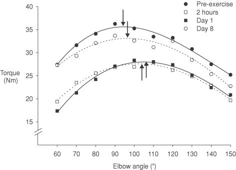
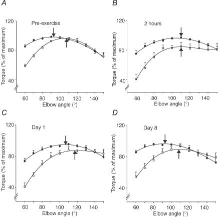
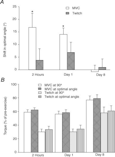
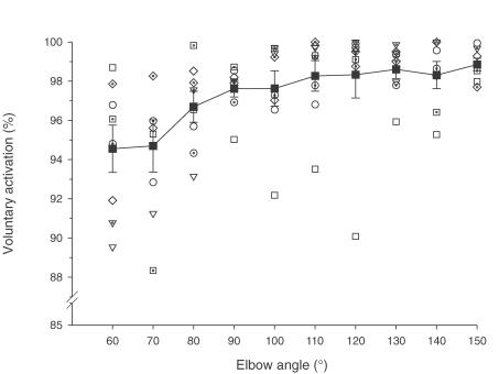
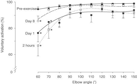
To assess the contribution of central and peripheral factors to changes in maximum voluntary force and its length dependence after eccentric muscle damage, voluntary and twitch torque were measured across a wide angular range, along with voluntary activation using twitch interpolation. Isometric torque from both maximum voluntary contractions (MVCs) and paired twitches to motor nerve stimulation were measured from 60 to 150 deg elbow flexion in 10 deg increments in eight subjects. Optimal angles were determined by curve fitting. Each subject then performed eccentric contractions until voluntary torque had decreased by approximately 40%. Measurements were repeated at 2 h, 1 day and 8 days post-exercise to follow acute and longer-term changes. Before exercise, the optimal angle was in the mid-range (93+/-10 deg; mean+/-s.d.) for MVCs, and at a more extended elbow angle for the twitch (106+/-6 deg, P < 0.05). Voluntary activation was generally high (> 94%) but depended on elbow angle, with activation being approximately 4% lower at the most flexed compared to the most extended angle. Two hours after exercise, MVCs decreased 40%, while twitch torque declined 70%. All subjects showed a shift in optimal angle to longer muscle lengths for MVCs (17+/-16 deg at 2 h, 14+/-7 deg at day 1, P < 0.05). This shift contributed minimally (approximately 3%) to the reduction in torque at 90 deg, as the torque-angle relation was relatively flat around the optimum. The twitch showed a smaller shift (approximately 4 deg) to longer lengths which was not statistically significant. Voluntary activation was significantly impaired in the early stages after exercise (2 h and day 1, P < 0.05), particularly at short muscle lengths. By 8 days after exercise, the optimal angle had returned to pre-exercise values, but MVC, twitch torque and voluntary activation had not fully recovered. Eccentric exercise causes a short-term shift in the optimal angle for MVCs and produces a length-dependent impairment in voluntary activation. Therefore, it appears that both central and peripheral factors limit muscle performance following eccentric damage, with limits to voluntary drive being especially important at short lengths.


Figures

Figure 1
Figure 1. Measurement protocols and twitch responses of elbow flexors in one subject
A, measurements of MVC, voluntary activation and twitch forces of elbow flexors were made at 60 deg elbow flexion and then the arm was passively extended in 10 deg steps in a custom-built myograph. Measurements were continued up to 150 deg and the sequence repeated. These sequences were performed pre-exercise, 2 h after eccentric exercise reduced the MVC by 40%, and 1 and 8 days later. Each shaded bar indicates the timing of voluntary and stimulated contractions and is expanded in panel B. B, during a MVC, paired stimuli (10 ms interval) were delivered to the motor nerve of biceps and brachialis. Paired and single stimuli were delivered after the MVC. C, resting twitches evoked by paired stimuli to the motor nerve for biceps and brachialis decreased markedly and gradually recovered by day 8. D, in this subject superimposed twitches evoked by the same stimuli during MVCs were similar in size before exercise, at day 1 and day 8.
Figure 2
Figure 2. Maximal voluntary isometric torque–angle curves from one subject
Data from one subject for MVCs at different elbow angles ranging from 60 deg to 150 deg before eccentric exercise (•), 2 h after (□), day 1 after (▪) and day 8 after (○). Data are the mean of two values obtained at each angle. Across the angular range, MVC was reduced at day 1 and recovered by day 8. The optimal muscle length at which the peak MVC occurred (vertical arrows) shifted acutely to a longer muscle length and returned close to the pre-exercise value at day 8.
Figure 3
Figure 3. Group data for maximal voluntary torques and twitches from paired stimuli before eccentric exercise, 2 h, 1 and 8 days following exercise
A, the optimal angles for MVC (•) and twitch from paired stimuli (□) before performing eccentric exercise were at 93 deg and 106 deg (arrows). Torques were normalized to their maximal values for each subject. Note that the ascending portion of the curves was steeper for the twitch than for the MVC at all time points. B, at 2 h after exercise, the optimal angles for the MVC and the twitch were at 110 deg. C, at day 1 after exercise, the optimal angle for the MVC and the twitch remained at the longer muscle length. D, at day 8 after exercise, the optimal angles for MVC and twitch were close to pre-exercise values. All data are shown as mean ±s.e.m. (n = 8).
Figure 4
Figure 4. Shift in the optimal angle for maximal voluntary torque and the twitch torque evoked by paired stimuli
A, over the first 24 h after eccentric exercise, the optimal angle shifted to a significantly longer length for MVC (open bars). The trend for a shift in the twitch was not significant (cross-hatched bars). Optimal angles were the same as pre-exercise values at day 8. B, voluntary torque and twitch evoked by paired stimuli at 90 deg and at the optimal angle. For the MVCs and twitches, there was no significant difference between torque at 90 deg and at the optimal angle at 2 h, day 1 and day 8. All data are shown as the mean ±s.e.m. (n = 8). Asterisks indicate a significant change from pre-exercise values.
Figure 5
Figure 5. Voluntary activation of elbow flexors before exercise for individual subjects and the group
Data for voluntary activation in MVCs for individual subjects obtained under control conditions are plotted across the angular range. Each open or dotted-open symbol shows data from an individual subject. Five of eight subjects had significantly lower voluntary activation at short lengths (see text). The mean for the group is also shown (filled squares with lines). At short lengths, voluntary activation was reduced for the group (P < 0.05).
Figure 6
Figure 6. Voluntary activation of elbow flexors across the angular range before and after eccentric exercise
Voluntary activation during MVCs before exercise (•) was high across the angular range (see Fig. 5). However, after exercise voluntary activation was impaired (P < 0.001), especially at short muscle lengths (60 deg and 70 deg) at 2 h (□) and at day 1 (▪), and only at 60 deg at day 8 (○). Data are shown as the mean ±s.e.m. (n = 8). Asterisks indicate a significant change from pre-exercise values at specific angles.

References

    1. An KN, Hui FC, Morrey BF, Linschied RL, Chao EY. Muscles across the elbow joint: a biomechanical analysis. J Biomech. 1981;14:659–669. - PubMed
    1. An KN, Kaufman KR, Chao EY. Physiological considerations of muscle force through the elbow joint. J Biomech. 1989;22:1249–1256. - PubMed
    1. Babault N, Pousson M, Michaut A, Van Hoecke J. Effect of quadriceps femoris muscle length on neural activation during isometric and concentric contractions. J Appl Physiol. 2003;94:983–990. - PubMed
    1. Balnave CD, Allen DG. Intracellular calcium and force in single mouse muscle fibres following repeated contractions with stretch. J Physiol. 1995;488:25–36. - PMC - PubMed
    1. Balnave CD, Allen DG. The effect of muscle length on intracellular calcium and force in single fibres from mouse skeletal. J Physiol. 1996;492:705–713. - PMC - PubMed

Publication types