. 2005 Dec 15;65(24):11493-500.
doi: 10.1158/0008-5472.CAN-05-2211.

Expression of endogenous oncogenic V600EB-raf induces proliferation and developmental defects in mice and transformation of primary fibroblasts

Affiliations

Expression of endogenous oncogenic V600EB-raf induces proliferation and developmental defects in mice and transformation of primary fibroblasts

Kathryn Mercer et al. Cancer Res. .

Abstract

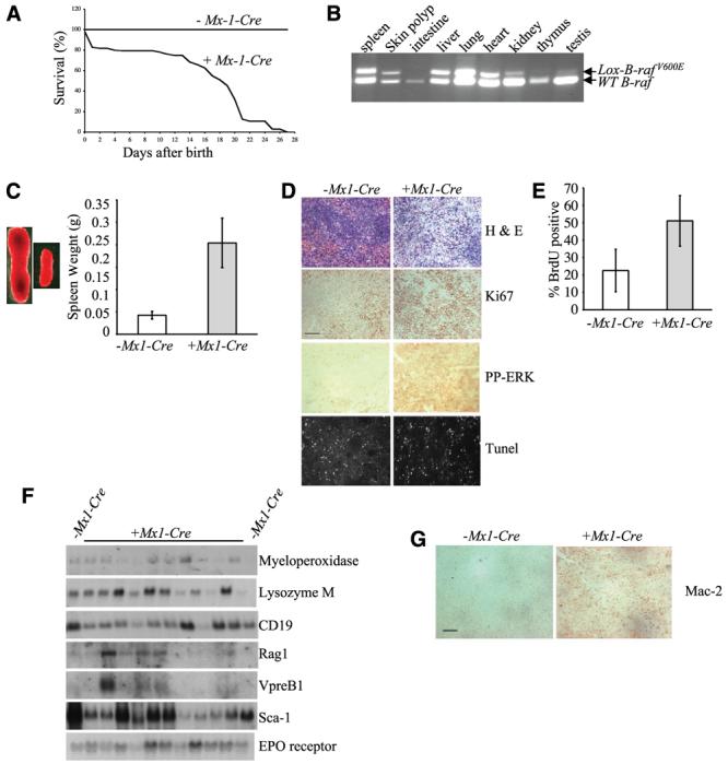
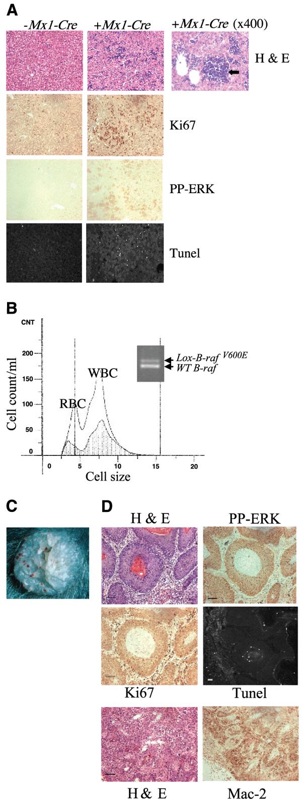
Mutations of the human B-RAF gene are detected in approximately 8% of cancer samples, primarily in cutaneous melanomas (70%). The most common mutation (90%) is a valine-to-glutamic acid mutation at residue 600 (V600E; formerly V599E according to previous nomenclature). Using a Cre/Lox approach, we have generated a conditional knock-in allele of (V600E)B-raf in mice. We show that widespread expression of (V600E)B-Raf cannot be tolerated in embryonic development, with embryos dying approximately 7.5 dpc. Directed expression of mutant (V600E)B-Raf to somatic tissues using the IFN-inducible Mx1-Cre mouse strain induces a proliferative disorder and bone marrow failure with evidence of nonlymphoid neoplasia of the histiocytic type leading to death within 4 weeks of age. However, expression of mutant B-Raf does not alter the proliferation profile of all somatic tissues. In primary mouse embryonic fibroblasts, expression of endogenous (V600E)B-Raf induces morphologic transformation, increased cell proliferation, and loss of contact inhibition. Thus, (V600E)B-Raf is able to induce several hallmarks of transformation in some primary mouse cells without evidence for the involvement of a cooperating oncogene or tumor suppressor gene.


Figures

Figure 1
Figure 1
A, generation of LSL-B-rafV600E and Lox-B-rafV600E alleles. The targeting vector contains left and right arms with the right arm containing exon 15 with the T1799A mutation (*). These two arms are separated by the LSL cassette. This cassette contains three LoxP sequences (black arrows), a minigene (MG) encoding exons 15 to 18 of wild-type B-Raf with a splice acceptor (SA) sequence at the 5′end. Two STOP sequences, represented by polyadenylation (PA) sequences, are located at the 3′end of the minigene and at the 3′end of the neoR cassette. Homologous recombination between the targeting vector and the wild-type B-raf gene in embryonic stem cells generated the LSL-B-rafV600E allele. Expression of the Cre recombinase allows deletion of the LSL cassette and generation of the Lox-B-rafV600E allele. Location of primers used for PCR genotyping (arrows and A-C). B, PCR genotyping to detect LSL-B-rafV600E, WT B-raf, and Cre alleles. Primers A-E were used in combination on tail DNA samples from intercrosses between heterozygous LSL-B-rafV600E mice and Cre mice. In this example, the WT B-raf allele (466 bp), Cre allele (350 bp), and LSL-B-rafV600E allele (140 bp) are analyzed in tail DNAs from heterozygous LSL-B-rafV600E mice without Cre (lane 1), wild-type mice with Cre (lane 2), and heterozygous LSL-B-rafV600E mice with Cre (lane 3). C, PCR genotyping to detect Lox-B-rafV600E and WT B-raf alleles. Primers A and C were used on the same DNA samples described above in (B). A 518-bp product indicates the presence of the Lox-B-rafV600E, and a 466-bp product indicates the presence of the WT B-raf allele. D, photographs of E7.5 embryos resulting from LSL-B-rafV600E × CMV-Cre intercrosses showing LSL-B-rafV600ECMV-Cre embryo (left) and LSL-B-rafV600E + CMV-Cre embryo (right). Bar, 250 μm.
Figure 2
Figure 2
Analysis of mice resulting from Lox-B-rafV600E × Mx1-Cre intercrosses. A, % survival of LSL-B-rafV600E + Mx1-Cre animals versus LSL-B-rafV600EMx1-Cre animals at various ages after birth. B, PCR genotyping to detect Lox-B-rafV600E allele in DNA from various tissues derived from LSL-B-rafV600E + Mx1-Cre mice. C, splenomegaly in LSL-B-rafV600E + Mx1-Cre mice. Left, increase in size of the spleen of P21 animals. Right, pooled weight data from four spleens of LSL-B-rafV600E ± Mx1-Cre mice at P21. LSL-B-rafV600E + Mx1-Cre mice have a mean spleen weight of 0.2 g compared with 0.04 g for the control LSL-B-rafV600EMx1-Cre mice. Columns, means; bars, SD. D, histologic analysis of spleen sections from LSL-B-rafV600E ± Mx1-Cre mice. Spleen sections were either stained with H&E, with antibodies for Ki67 or phospho-ERK, or processed for TUNEL analysis. Bar, 50 μm (the same for all sections). E, proliferation analysis of spleens from LSL-B-rafV600E ± Mx1-Cre mice. Mice were injected with BrdUrd, splenocytes were isolated, and the % incorporation of BrdUrd was assessed by FACS analysis. Columns, means; bars, SD. F, Northern blot analysis of spleen RNA isolated from LSL-B-rafV600E ± Mx1-Cre mice. Northern blots were probed with the markers indicated. G, histologic analysis of spleen sections from LSL-B-rafV600E ± Mx1-Cre mice with a histiocyte marker. Spleen sections were stained with a Mac-2 antibody to detect histiocyte amplification. Bar, 50 μm.
Figure 3
Figure 3
Analysis of tissues from mice resulting from LSL-B-rafV600E × Mx1-Cre intercrosses. A, histologic analysis of liver sections from LSL-B-rafV600E ± Mx1-Cre mice. Liver sections were either stained with H&E, with a Ki67 antibody, with a phospho-ERK antibody or processed for TUNEL analysis. Right, H&E section was photographed at higher magnification (×400) to indicate site of hemopoiesis (black arrow) in the mutant liver. Bar, 50 μm (the same for all sections) except for 25 μm (enlarged H&E section, right). B, analysis of bone marrow from LSL-B-rafV600E ± Mx1-Cre mice. Bone marrow cells were isolated from the femurs of LSL-B-rafV600E ± Mx1-Cre mice and analyzed by the Coulter counter. There are far fewer RBC and WBC in the bone marrow of LSL-B-rafV600E + Mx1-Cre mice (shaded area) than LSL-B-rafV600EMx1-Cre mice (clear area). PCR analysis of DNA from bone marrow of LSL-B-rafV600E + Mx1-Cre animal, indicating the presence of the Lox-B-rafV600E allele. C, photograph of histiocyte arising on skin of LSL-B-rafV600E + Mx1-Cre animal. D, histologic analysis of histiocyte sections from LSL-B-rafV600E ± Mx1-Cre mice. Sections were either stained with H&E, with antibodies for Ki67, phospho-ERK, or Mac-2, or processed for TUNEL analysis. Bar, 50 μm.
Figure 4
Figure 4
Analysis of B-RAF/MEK/ERK activation in tissues derived from LSL-B-rafV600E ± Mx1-Cre mice. Protein lysates were harvested from various tissues and subjected to (A) B-Raf kinase assays and (B) Western blot analysis with antibodies for phospho-ERK, phospho-MEK, and cyclin D1. Immunoblotting with a total ERK antibody was used to confirm protein loading.
Figure 5
Figure 5
Analysis of primary MEFs derived from LSL-B-rafV600E mice. A, morphologic transformation. Primary LSL-B-rafV600E MEFs were either mock-transfected (−Cre) or transfected with the pCrePac plasmid (+Cre). After 48 hours, the cells were either photographed (top) or subjected to immunofluorescence analysis with FITC-phalloidin (bottom). B, proliferation analysis. Mock- and Cre-transfected cells were serum starved for 20 hours (open columns) and then stimulated with serum for 16 hours (gray columns). % BrdUrd incorporation was assessed under each condition. Columns, means; bars, SD. The cells transfected with Cre undergo a high level of proliferation even in the absence of serum compared with mock-transfected controls. C, Western blot analysis of lysates prepared from Cre- and mock-transfected MEFs with an antibody for phospho-ERK. Immunoblotting with a total ERK antibody was used to confirm protein loading. D, Western blot analysis of lysates prepared from Cre- and mock-transfected MEFs with antibodies for cyclin D1 and p21CIP1. Immunoblotting with an antibody for actin was used to confirm protein loading. E, focus formation assay of Cre- and mock-transfected LSL-B-rafV600E primary MEFs. MEFs were grown to confluency and the media was changed every 2 to 3 days until foci formed for 18 days. F, soft agar assays of Cre- and mock-transfected LSL-B-rafV600E primary MEFs.

References

    1. Kan NC, Flordellis CS, Garon CF, Duesberg PH, Papas TS. Avian carcinoma virus MH2 contains a transformation-specific sequence, mht, and shares the myc sequence with MC29, CMII, OK10 viruses. Proc Natl Acad Sci U S A. 1983;80:6566–70. - PMC - PubMed
    1. Rapp UR, Goldsborough MD, Mark GE, et al. Structure and biological activity of v-raf, a unique oncogene transduced by a retrovirus. Proc Natl Acad Sci U S A. 1983;80:4218–22. - PMC - PubMed
    1. Jansen HW, Lurz R, Bister K, Bonner TI, Mark GE, Rapp UR. Homologous cell-derived oncogenes in avian carcinoma virus MH2 and murine sarcoma virus 3611. Nature. 1984;307:281–4. - PubMed
    1. Storm SM, Brennscheidt U, Sithanandam G, Rapp UR. raf oncogenes in carcinogenesis. Crit Rev Oncog. 1990;2:1–8. - PubMed
    1. Davies H, Bignell GR, Cox C, et al. Mutations of the BRAF gene in human cancer. Nature. 2002;417:949–54. - PubMed

Publication types

MeSH terms