. 2005 Dec 27;102(52):18986-91.
doi: 10.1073/pnas.0509535102. Epub 2005 Dec 15.

MicroRNA1 influences cardiac differentiation in Drosophila and regulates Notch signaling

Affiliations

MicroRNA1 influences cardiac differentiation in Drosophila and regulates Notch signaling

Chulan Kwon et al. Proc Natl Acad Sci U S A. .

Abstract

MicroRNAs (miRNAs) are genomically encoded small RNAs that hybridize with messenger RNAs, resulting in degradation or translational inhibition of targeted transcripts. The potential for miRNAs to regulate cell-lineage determination or differentiation from pluripotent progenitor or stem cells is unknown. Here, we show that microRNA1 (miR-1) is an ancient muscle-specific gene conserved in sequence and expression in Drosophila. Drosophila miR-1 (dmiR-1) is regulated through a serum response factor-like binding site in cardiac progenitor cells. Loss- and gain-of-function studies demonstrated a role for dmiR-1 in modulating cardiogenesis and in maintenance of muscle-gene expression. We provide in vivo evidence that dmiR-1 targets transcripts encoding the Notch ligand Delta, providing a potential mechanism for the expansion of cardiac and muscle progenitor cells and failure of progenitor cell differentiation in some dmiR-1 mutants. These findings demonstrate that dmiR-1 may "fine-tune" critical steps involved in differentiation of cardiac and somatic muscle progenitors and targets a pathway required for progenitor cell specification and asymmetric cell division.


Figures

Fig. 1.
Fig. 1.
dmiR-1 transcripts mark mesodermal cells and derivatives. (a–f) Whole-mount in situ hybridization of miR-1 (blue) at stages 5 (a),9(b), 11(c), 13 (d), and 16 (e and f) embryos. dmiR-1 is expressed in the presumptive mesoderm (a), mesodermal cells (b–d) and derivatives such as the heart tube (arrowhead in e), and somatic and visceral muscles (arrows in e and f, respectively). Embryos in a–d were costained with anti-Dmef2 (red). (a–f) Anterior left, posterior right views; (a–d) lateral views; (e) dorsolateral view; (f) interior confocal view of e.
Fig. 2.
Fig. 2.
Regulation of dmiR-1 in cardioblast and visceral muscle cells. (a) Map of the dmiR-1 locus showing the position of the 4.6-kb dmiR-1 enhancer (green) and subfragments, with expression domains summarized as follows: SM, somatic muscle; VM, visceral muscle; CB, cardioblast; PC, pericardial cell. An A/T-rich SRF-like-binding site conserved in other Drosophila species is highlighted. (b–m) GFP expression in embryos carrying the 4.6-kb (b–g), 2.5-kb (h and i), 0.72-kb (j and l), or SRF-like site-mutated 0.72-kb (k and m) element. Embryos were costained with anti-Dmef2 (c and i) or anti-Twist (e and g). (n) Luciferase (luc) activity determined with luciferase reporters linked to the 0.72-kb element or the SRF-like site mutated (CArGm) 0.72-kb element in Drosophila S2 cells in the presence or absence of Drosophila SRF and myocardin-related transcription factor. Error bars indicate standard deviations. (b, c, h–k) Dorsolateral views of stage 16 embryos. (d–g) Lateral views of stage 11 embryos. f and g are ×40 images of d and e, respectively. l and m are inside views of j and k embryos, respectively, focusing on visceral muscles. Arrowheads indicate the presence (b, h, and j) or absence (k) of the heart tube, and arrows indicate somatic (b, h, k, and j) and visceral muscles (l).
Fig. 3.
Fig. 3.
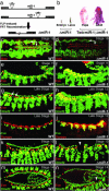
Loss of dmiR-1 causes abnormal heart and muscle development. (a) Schematic of FRT-FLP-mediated dmiR-1 locus deletion and locations of neighboring genes. Embryonic and larval lethality was rescued by UAS-miR-1 with twi-gal4 driver (b) or by a transgene containing the 5.1-kb miR-1 locus (not shown). (c, e, g, i, k, and m) WT embryos. (d, f, h, j, l, and n) Homozygous miR-1 mutant (ΔmiR-1) embryos. Expression of MHC (myosin heavy chain) was dramatically reduced both in the heart and muscles (arrows in c and d) in half of all ΔmiR-1 embryos compared with WT, whereas expression of a pericardial cell marker, odd-skipped, was not affected (c and d). ΔmiR-1 embryos with the early defect had ectopic Eve progenitor cells at stage 11 (f). The ectopic progenitor cells failed to differentiate into pericardial (arrows in g) and dorsal muscles (asterisks) and maintained expression of both Dmef2 and eve at late-stage 12 (h). Ectopic Dmef2+ cardioblasts, identified based on their dorsal location relative to the Eve progenitors, were also observed (h). tinman was expressed in most of the ectopic cardiac progenitors that expressed eve (j). (k–n) Another subset of embryos that fail to hatch showed reduced numbers of cardiac and muscle cells, indicated by gaps in the row of cardioblasts and missing dorsal muscles in ΔmiR-1 embryos (arrowheads in l and n). (e–l) Lateral views; (c, d, m, and n) dorsal views.
Fig. 4.
Fig. 4.
dmiR-1 overexpression in the mesoderm affects heart and muscle cell morphology. (a–d) Lateral views of stage 13 WT (a and b) or dmiR-1 overexpressed embryos with 24B-Gal4 driver (c and d). b and d are ×40 images of white boxes in a and c, respectively. On average, four rather than six cardioblasts per hemisegment were observed in transgenic flies with occasional enlarged cardioblasts (arrowhead).
Fig. 5.
Fig. 5.
dmiR-1 can target the Notch ligand Delta. (a) Relative luciferase activity was determined by inserting part of the 3′-UTR target sequence from Delta, recognized by dmiR-1, into the 3′-UTR of luciferase (Delta 3′-UTR-luc) in the presence or absence of dmiR-1 in Drosophila S2 cells. The basal level of Delta 3′-UTR-luc or Delta 3′-UTR-mut-luc activity in the presence of Ubi-Gal4 but not UAS-miR-1 was defined as 100%. Overexpression of dmiR-1 inhibited activity of the luciferase reporter with WT Delta 3′UTR, but not with the dmiR–binding-site mutated Delta 3′UTR. Error bars indicate standard deviation. (b) Delta expression in the WT wing pouch using anti-Delta antibody (red). (c) Ectopic dmiR-1 expression in the dpp expression domain (indicated by a yellow stripe in the wing pouch cartoon in c) resulted in severely compromised endogenous Delta protein expression along the dmiR-1-overexpressed domain. (d) WT adult wing and leg. (e) Overexpression of dmiR-1 in wing and leg discs caused thickened wing veins (arrows) specifically in longitudinal veins 3 and 4 (L3 and L4) and shortened legs, similar to the loss of Delta phenotype. (f) Hypothetical model for early function of dmiR-1 in mesodermal cells through regulation of Notch signaling. dmiR-1 may be important in reinforcing selection of lineages from equivalency groups normally regulated by Notch. Disruption of the normal segregation of Delta- and Notch-expressing cells in the absence of dmiR-1 may result in excess Delta-expressing cells and consequent down-regulation of Notch and failure of lineage determination. L1–5, longitudinal vein 1–5; Fe, femur; Ti, tibia; Ta1–5, tarsal segment 1–5; Cl, claw.

References

    1. Ambros, V. (2001) Cell 107, 823–826. - PubMed
    1. Johnston, R. J. & Hobert, O. (2003) Nature 426, 845–849. - PubMed
    1. Bartel, D. P. (2004) Cell 116, 281–297. - PubMed
    1. Lewis, B. P., Shih, I. H., Jones-Rhoades, M. W., Bartel, D. P. & Burge, C. B. (2003) Cell 115, 787–798. - PubMed
    1. Zhao, Y., Samal, E. & Srivastava, D. (2005) Nature 436, 214–220. - PubMed

Publication types

MeSH terms

LinkOut - more resources