. 2005 Dec 27;102(52):18902-7.
doi: 10.1073/pnas.0509069102. Epub 2005 Dec 15.

A therapeutic aptamer inhibits angiogenesis by specifically targeting the heparin binding domain of VEGF165

Affiliations

A therapeutic aptamer inhibits angiogenesis by specifically targeting the heparin binding domain of VEGF165

Joon-Hwa Lee et al. Proc Natl Acad Sci U S A. .

Abstract

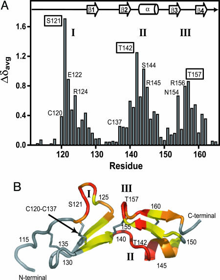
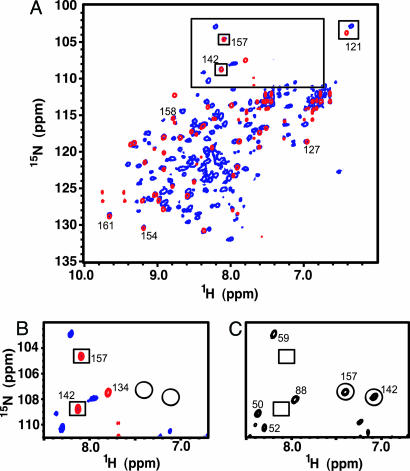
Aptamers recognize their targets with extraordinary affinity and specificity. The aptamer-based therapeutic, Macugen, is derived from a modified 2'fluoro pyrimidine RNA inhibitor to vascular endothelial growth factor (VEGF) and is now being used to treat the wet form of age-related macular degeneration. This VEGF(165) aptamer binds specifically to the VEGF(165) isoform, a dimeric protein with a receptor-binding domain and a heparin-binding domain (HBD). To understand the molecular recognition between VEGF and this aptamer, binding experiments were used to show that the HBD contributes the majority of binding energy in the VEGF(165)-aptamer complex. A tissue culture-based competition assay demonstrated that the HBD effectively competes with VEGF(165) for aptamer binding in vivo. Comparison of NMR spectra revealed that structural features of the smaller HBD-aptamer complex are present in the full-length VEGF(164)-aptamer complex. These data show that the HBD provides the binding site for the aptamer and is the primary determinant for the affinity and specificity in the VEGF(165)-aptamer complex.


Figures

Fig. 1.
Fig. 1.
The HBD competes with VEGF165 for aptamer binding in vivo. (A) Sequence and secondary structure of the VEGF aptamer. The subscripts m and f indicate the 2′-OMethyl- and 2′-fluoro-modified residues, respectively, and A represents unmodified ribo-A. The arrow points to the photo-crosslinking site at U14. (B) In vivo competition binding assay for the HBD and VEGF165. Relative mRNA expression levels of TF in HUVEC in culture are normalized to untreated cells. Increasing amounts of HBD (2.3, 4.7, 9.4, 18.8, 37.5, 75, 150, and 300 nM) were added to HUVEC treated with VEGF165 and aptamer. The data show that addition of HBD can effectively reverse the inhibition of VEGF165-induced TF expression by the aptamer. Controls include VEGF165- or VEGF121-induced cells, untreated cells, or addition of DTT-inactivated HBD.
Fig. 2.
Fig. 2.
Characteristic NMR fingerprint of aptamer in complex with the HBD and VEGF164. (A) Imino proton spectra of the aptamer at 10°C upon titration with HBD. The molar ratios are shown on the left. (B) Comparison of the imino proton spectra of the aptamer at 40°C. (Top) The free aptamer. (Middle) The HBD–aptamer complex. (Bottom) The VEGF164–aptamer complex. (C) Comparison of the 31P NMR spectra of the aptamer. (Top) The free aptamer at 25°C. (Middle) The HBD–aptamer complex at 25°C. (Bottom) The VEGF164–aptamer complex at 45°C.
Fig. 3.
Fig. 3.
Superposition of 1H-15N HSQC spectra at 10°C of free HBD (blue) and aptamer-bound HBD (red). Backbone resonance assignments are indicated in blue (free HBD), red (HBD–aptamer complex), and black (free and bound HBD). Boxes illustrate the NMR fingerprint created by residues S121, T142, and T157 in the HBD–aptamer complex.
Fig. 4.
Fig. 4.
HBD chemical shift changes upon aptamer binding. (A) The weighted average 1H/15N backbone amide chemical shift changes (Δδavg) of the HBD upon binding to the aptamer. Three regions with large chemical shift changes are labeled as I, II, and III. The residues with the largest chemical shift changes in each region are boxed (S121, T142, and T157). The positions of the secondary structure elements are shown for the free HBD (6). (B) Secondary structural model for the previously determined NMR structure of the free HBD (6). Colors used to illustrate 1H/15N backbone chemical shift changes (Δδavg) upon aptamer binding are: red, >0.5 ppm; magenta, 0.3–0.5 ppm; yellow, 0.1–0.3 ppm; gray, <0.1 ppm. The three regions with large chemical shift changes are labeled I, II, and III.
Fig. 5.
Fig. 5.
Protein NMR fingerprint of HBD–aptamer and VEGF164–aptamer complexes. (A) Superposition of the 1H-15N TROSY-HSQC spectra of the HBD–aptamer complex (red) and VEGF164–aptamer complex (blue) acquired at 35°C at 800 MHz. The characteristic signature created by S121, T142, and T157 is emphasized (boxes). (B) Expansion of the spectral region containing the T142 and T157 crosspeaks in the aptamer-bound spectra of the HBD (red) and VEGF164 (blue). The circles indicate the position of these crosspeaks in the free HBD and free VEGF164. (C) The same spectral region as B showing the 1H-15N TROSY-HSQC spectra of free VEGF164. The T142 and T157 amide crosspeaks are circled; the boxes indicate the positions of these crosspeaks in the VEGF164–aptamer complex.

References

    1. Ferrara, N., Gerber, H. P. & LeCouter, J. (2003) Nat. Med. 9, 669–676. - PubMed
    1. Tammela, T., Enholm, B., Alitola, K. & Paavonen, K. (2005) Cardiovasc. Res. 65, 550–553. - PubMed
    1. Witmer, A. N., Vrensen, G., Van Noorden, C. J. F. & Schlingemann, R. O. (2003) Prog. Retin. Eye Res. 22, 1–29. - PubMed
    1. Ishida, S., Usui, T., Yamashiro, K., Kaji, Y., Amano, S., Ogura, Y., Hida, T., Oguchi, Y., Ambati, J., Miller, J. W., et al. (2003) J. Exp. Med. 198, 483–489. - PMC - PubMed
    1. Ng, E. & Adamis, A. (2005) Can. J. Ophthalmol. 40, 352–368. - PubMed

Publication types

MeSH terms