. 2005 Dec 15;19(24):3004-16.
doi: 10.1101/gad.353205.

The RNA polymerase II subunit Rpb4p mediates decay of a specific class of mRNAs

Affiliations

The RNA polymerase II subunit Rpb4p mediates decay of a specific class of mRNAs

Rona Lotan et al. Genes Dev. .

Abstract

It is commonly appreciated that the mRNA level is determined by the balance between its synthetic and decay kinetics. Yet, little is known about coordination between these distinct processes. A major pathway of the eukaryotic mRNA decay initiates with shortening of the mRNA poly(A) tail (deadenylation), followed by removal of the mRNA 5' cap structure and its subsequent exonucleolytic degradation. Here we report that a subunit of RNA polymerase II, Rpb4p, is required for the decay of a class of mRNAs whose products are involved in protein synthesis. Cells lacking RPB4 are defective in the deadenylation and post-deadenylation steps of representatives of this class of mRNAs. Moreover, Rpb4p interacts with both the mRNP and with subunits of the mRNA decay complex Pat1/Lsm1-7 that enhances decapping. Consistently, a portion of Rpb4p is localized in P bodies, where mRNA decapping and degradation is executed, and mutations in RPB4 increase the number of P bodies per cell. We propose that Rpb4p has a dual function in mRNA decay. It promotes or enhances the deadenylation process of specific mRNAs and recruits Pat1/Lsm1-7 to these mRNAs, thus stimulating their decapping and further decay. In this way, Rpb4p might link the activity of the basal transcription apparatus with that of the mRNA decay machinery.


Figures

Figure 1.
Figure 1.
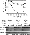
Rpb4p is required for efficient decay of RPL25 and NSR1 mRNAs at both 24°C and 37°C. Wild-type (WT) strain (SUB62) and its isogenic rpb4Δ derivative (MC11-1) were proliferated at 24°C until mid-logarithmic phase. Transcription was then blocked by 1, 10 phenanthroline and the cell culture was then incubated at either 24°C (A) or 37°C (B). Decay kinetics was then determined by monitoring mRNA levels at the indicated time points post-drug treatment, using Northern analysis (as detailed in Materials and Methods). The filter was reacted sequentially with the probes that are indicated at the left in A. rRNA (18S) is shown to demonstrate equal loading. Decreased kinetics of mRNA level was determined by PhosphorImager. Band intensity at time 0 (before adding the drug) was defined as 1 and the intensities at the other time points were calculated relative to time 0. Results were plotted as a function of time post-drug addition. This analysis was used to determine half-lives (designated T½). The upper panels show the results of RPL25 mRNA decay kinetics.
Figure 2.
Figure 2.
Rpb4p's role in the mRNA decay pathway is specific. (A) Degradation kinetics was determined in wild-type (WT) and rpb4Δ strains as in Figure 1 (37°C). The filter was hybridized sequentially to the indicated probes. Half-lives were determined as in Figure 1, and the ratios between T½ of the mutant and that of the wild-type [T½ (m)/T½ (WT)] are indicated on the right. rRNA (18S) is shown to demonstrate equal loading. Note that the identical decay kinetics of MFA2, ACT1, and TDH3 mRNAs can serve also as an internal control for equal loading. (B) Decay kinetics of ACT1 mRNA in the wild-type (diamonds)and in rpb4Δ (squares) were determined as in Figure 1.
Figure 3.
Figure 3.
Overexpression of RPB7 does not change the defective decay kinetics of RPL25 and RPL28 mRNAs in rpb4Δ cells. Degradation kinetics in three isogenic strains (see Materials and Methods) was determined as in Figure 1. The absence of a gene (-), the presence of one copy of a gene (+), and the overexpression of a gene (+++) are indicated.
Figure 4.
Figure 4.
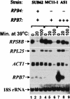
Rpb4p's role in mRNA decay is displayed after exposing cells to heat shock. Cells of the indicated strains (see Materials and Methods) were allowed to proliferate in galactose-containing medium (to permit overexpression of pGAL1p-RPB7) at 24°C until mid-logarithmic phase. Cell aliquots were taken for time 0 and then the cultures were shifted to 39°C. Cell samples were harvested at the indicated time points after the shift. Northern analysis was done as in Figure 1. The absence of a gene (-), the presence of one copy of a gene (+), and the overexpression of a gene (+++) are indicated.
Figure 5.
Figure 5.
Rpb4p is required for efficient deadenylation and subsequent decay of RPL29 mRNA, but not of MFA2 mRNA. Cells of the indicated strains were allowed to proliferate at 24°C until mid-logarithmic phase. Cell aliquots were taken for time 0 and then the culture was shifted rapidly to 42°C to arrest transcription of PBF genes. (A) Cell samples were harvested at the indicated time points after the shift and their RNA were analyzed by the polyacylamide Northern technique (see Materials and Methods) and probed first with RPL29. (B) The membrane was then stripped off the first probe and hybridized with MFA2 probe. Lane “Δ(A)n” shows the position of fully deadenylated RNA. This RNA was obtained by hybridizing the RNA sample of time point 0 with oligo(dT) followed by digestion of the poly(A) tail by RNase H. Positions of single-stranded DNA size marker are indicated at the right.
Figure 6.
Figure 6.
Rpb4p is not essential for NMD. Two iogenic pairs were harvested in mid-logarithmic growth phase and the steadystate levels of their RPL28 (CHY2) mRNAs were analyzed by Northern anaylsis as in Figure 1. The probe was specific for both the intron and the coding region of RPL28, spanning positions 24–730 from ATG in the genomic sequence. Marked by arrows are the positions of RPL28 pre-mRNA (unspliced) and the mature (spliced) mRNA in upf1Δ and its isogenic wild-type (WT) strain (4741) (left lanes) and rpb4Δ and its isogenic wild-type strain (SUB62) (right lanes). The lane second from the right is empty. EtBr stained 18S rRNA is shown at the bottom to demonstrate that each isogenic pair was loaded equally.
Figure 7.
Figure 7.
Rpb4p is colocalized to P bodies and affects the P bodies' function. (A) Colocalization of Rpb4p with the P bodies' marker. Cells expressing the indicated tagged proteins were allowed to proliferate in selective medium at 24°C. (Upper panels) Cells in mid-log phase were visualized by fluorescent microscopy at either the green (left) or the red (middle) channel. The right panels show the merge generated by Adobe Photoshop. Cells were then shifted to starvation medium lacking any sugar and lacking amino acids for 4 h (middle panels) or 24 h (lower panels). Arrows in the upper panels point at some GFP-Rpb4p containing foci, found rarely in optimally growing cells or after 4 h of starvation. (B) Colocalization of Rpb4–26p with the P bodies' markers. Cells expressing GFP-rpb4–26p, in lieu of RPB4, and Lsm1p-RFP were allowed to proliferate until mid-log followed by 4 h starvation period, as in the middle panels in A. Cells were visualized by fluorescence microscopy (upper panels) or by confocal microscopy (bottom panels). It is worth noting that Rpb4–26-RFP, like GFP-Rpb4–26p, was localized in discrete foci (results not shown), indicating that this type of localization is not a GFP-specific artifact. (C) RPB4 affects the P bodies' number. Wild-type (WT) cells (YMC274) and their isogenic rpb4Δ derivatives (YCM275), both expressing LSM1-RFP, were allowed to proliferate in a selective medium until early stationary phase (24 h post-diauxic shift), before they were visualized by fluorescent microscopy to detect Lsm1p-RFP-containing P bodies. (D) Effect of cycloheximide on GFP-Rpb4-containing foci. Cells expressing GFP-rpb4–26, in lieu of RPB4, were allowed to proliferate in synthetic medium at 24°C. When the culture approached mid-log, it was divided into two samples: one treated with 100 μM cycloheximide (CHX) and the other with the drug vehicle (ethanol) (Cont.). Cells were incubated with the drug for 15 min at 24°C and then shifted to the starvation medium lacking a carbon source and amino acids, which included the drug or the drug vehicle. GFP-Rpb4–26p localization was determined 4 h later.
Figure 8.
Figure 8.
RPB4 interacts with PAT1 and LSM2. (A) Physical interactions. (B) Genetic interactions. (C) Two-hybrid interactions. (A) Coimmunoprecipitation of Rpb4 that was tagged with “tandem affinity purification” (TAP) (Gavin et al. 2002) (designated Rpb4-TAP) and Pat1p and Npl3p in the presence or absence of RNase A. Nucleoproteins were extracted from the strain expressing either RPB3-TAP (as the control) or RPB4-TAP under conditions that maintained intact RNAs (see Materials and Methods). Immunoprecipitation with IgG sepharose was done from the indicated extracts with or without prior RNase A treatment (indicated above the lanes), and the various proteins present in the extract (designated Ext) or in the flow-through sample (designated FT) or in the immunoprecipitates were revealed by Western analysis as described in Materials and Methods. Rpb4-TAP (Rpb4-TAP shown in lanes 1,2, or its product released from the column by TEV protease digestion [Gavin et al. 2002], shown in lanes 4,5) was detected by anti-TAP antibodies (Open biosystem). These antibodies detected the Rpb3-CBD in lane 1 (not shown). Rpb7p that is coimmunoprecipitated with both Rpb4-TAP and Rpb3-TAP is shown to demonstrate equal immunoprecipitation efficiencies. (B) High copy suppression of pat1Δ defect by RPB4. Isogenic strains wild-type (lane 1), pat1Δ (lane 2), or pat1Δ + high-copy plasmid (pRS426 derivative) expressing RPB4 (lane 3) were spotted onto two plates in fivefold serial dilutions. Cells were allowed to grow at either 24°C or 36°C, as indicated. (C) Two-hybrid interactions between RPB4 as the bait and genes whose products are involved in mRNA decay as prey have been performed as described in Materials and Methods. Only five of 20 genes tested are shown as indicated around the plate. The other genes that did not exhibit interactions with the RPB4-DBD and are not shown here are LSM1, LSM4–LSM7, CCR4, NOT1, CAF1, PAN3, PAN2, POP2, EDC1, EDC2, EDC3, PAB1. Equal amount of cells, carrying the indicated prey plasmids, were streaked onto an indicator plate as described in Materials and Methods. We then verified that the growth on the indicator plates was dependent on both plasmids by evicting one plasmid at a time from each of the positive clones (results not shown). Units of β-gal, a third reporter of the two-hybrid interaction, were also determined by liquid tests and yielded results that were consistent with the growth phenotype (data not shown).

Similar articles

Cited by

References

    1. Armache K.J., Kettenberger, H., and Cramer, P. 2003. Architecture of initiation-competent 12-subunit RNA polymerase II. Proc. Natl. Acad. Sci. 100: 6964-6968. - PMC - PubMed
    1. Badis G., Saveanu, C., Fromont-Racine, M., and Jacquier, A. 2004. Targeted mRNA degradation by deadenylation-independent decapping. Mol. Cell 15: 5-15. - PubMed
    1. Bashkirov V.I., Scherthan, H., Solinger, J.A., Buerstedde, J.M., and Heyer, W.D. 1997. A mouse cytoplasmic exoribonuclease (mXRN1p) with preference for G4 tetraplex substrates. J. Cell. Biol. 136: 761-773. - PMC - PubMed
    1. Bonnerot C., Boeck, R., and Lapeyre, B. 2000. The two proteins Pat1p (Mrt1p) and Spb8p interact in vivo, are required for mRNA decay, and are functionally linked to Pab1p. Mol. Cell. Biol. 20: 5939-5946. - PMC - PubMed
    1. Brown C.E. and Sachs, A.B. 1998. Poly(A) tail length control in Saccharomyces cerevisiae occurs by message-specific deadenylation. Mol. Cell. Biol. 18: 6548-6559. - PMC - PubMed

Publication types

MeSH terms

Substances