. 2005 Dec 15;19(24):3043-54.
doi: 10.1101/gad.1373705.

The Rad50S allele promotes ATM-dependent DNA damage responses and suppresses ATM deficiency: implications for the Mre11 complex as a DNA damage sensor

Affiliations

The Rad50S allele promotes ATM-dependent DNA damage responses and suppresses ATM deficiency: implications for the Mre11 complex as a DNA damage sensor

Monica Morales et al. Genes Dev. .

Abstract

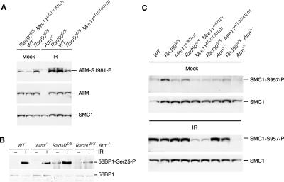
Genetic and cytologic data from Saccharomyces cerevisiae and mammals implicate the Mre11 complex, consisting of Mre11, Rad50, and Nbs1, as a sensor of DNA damage, and indicate that the complex influences the activity of ataxia-telangiectasia mutated (ATM) in the DNA damage response. Rad50(S/S) mice exhibit precipitous apoptotic attrition of hematopoietic cells. We generated ATM- and Chk2-deficient Rad50(S/S) mice and found that Rad50(S/S) cellular attrition was strongly ATM and Chk2 dependent. The hypomorphic Mre11(ATLD1) and Nbs1(Delta)(B) alleles conferred similar rescue of Rad50(S/S)-dependent hematopoietic failure. These data indicate that the Mre11 complex activates an ATM-Chk2-dependent apoptotic pathway. We find that apoptosis and cell cycle checkpoint activation are parallel outcomes of the Mre11 complex-ATM pathway. Conversely, the Rad50(S) mutation mitigated several phenotypic features of ATM deficiency. We propose that the Rad50(S) allele is hypermorphic for DNA damage signaling, and that the resulting constitutive low-level activation of the DNA damage response accounts for the partial suppression of ATM deficiency in Rad50(S/S) Atm(-/-) mice.


Figures

Figure 1.
Figure 1.
Rescue of Rad50S/S survival by Rad50-null, Atm-null, Mre11ATLD1, and Nbs1ΔB alleles. (A) Kaplan-Meier survival curves of Rad50S/S and Rad50Δ/S mice. (B) Kaplan-Meier survival curves of Rad50S/S, Atm-/-, Rad50S/S Atm+/-, and Rad50S/S Atm-/- mice. (C) Kaplan-Meier survival curves of Rad50S/S, Rad50S/S Mre11+/ATLD1, Rad50S/S Mre11ATLD/ATLD1, Rad50S/S Nbs1+/ΔB, and Rad50S/S Nbs1ΔB/ΔB mice. (AC) In each table below the survival curves, for each cohort, the number of mice that were found dead without overt signs of anemia and malignancy is equal to the number of mice minus the number of alive mice, the number of dead mice with anemia, and the number of dead mice with lymphoma. (D) Hematoxylin and eosin staining (upper pictures) or cleaved Caspase-3 detected by immunohistochemistry (lower pictures) in bone marrow sections from 4-wk-old Rad50S/S, Rad50S/S Atm-/-, Rad50S/S Mre11+/ATLD1, and Rad50S/S Mre11ATLD/ATLD1 mice. Magnification, 100×. Bar, 100 μM.
Figure 2.
Figure 2.
Rescue of Rad50S/S hematopoietic attrition by Atm-null, Mre11ATLD1, and Nbs1ΔB alleles, and by Chk2-/-. Flow cytometric analysis of hematopoietic tissues from 4–9-wk-old mice. The numbers of double-negative T cells in the thymus (DN T cells; A), and macrophages (B) and pro-B cells in the bone marrow (C), normalized to wild-type data, are depicted for the indicated genotypes.
Figure 3.
Figure 3.
Rescue of Rad50S/S survival by Chk2-null allele and the Eμ-Bcl2 transgene. (A) Kaplan-Meier survival curves of Rad50S/S and Rad50S/S Eμ-Bcl2 mice. (B) Kaplan-Meier survival curves of Rad50S/S, Rad50S/S Chk2+/-, Rad50S/S Chk2-/-, and Rad50S/S Smc12SA/2SA mice. Each table below the survival curves is as in Figure 1.
Figure 4.
Figure 4.
Constitutive ATM activity in Rad50S/S is rescued by ATM deficiency and Mre11ATLD1. (A) Analysis of ATM Ser1981 phosphorylation. Extracts from wild-type (WT), Rad50S/S, Rad50S/S Mre11ATLD1/ATLD1, and Atm-/- p4 ear fibroblasts were prepared after mock treatment and 1 h after 10 Gy of IR. For the ATM Ser1981 phosphorylation Western blot (ATM-Ser1981-P), the lower reactive species represents ATM, as indicated by the ATM Western blot (ATM), and the upper band is nonspecific. SMC1 is included as a loading control. (B) Analysis of 53BP1 phosphorylation in primary ear fibroblasts. Ser25-phosphorylated 53BP1 was immunoprecipitated from untreated or irradiated (10 min after 4 Gy) primary ear fibroblast extracts, and detected with a 53BP1 antibody. A 53BP1 Western blot of the same extracts is shown as a loading control. (C) Analysis of SMC1 Ser 957 phosphorylation in the indicated mutants. Extracts from p4 ear fibroblasts were prepared after mock treatment and 1 h after 10 Gy of IR. The extracts were sequentially immunoblotted with SMC1-Ser957-P and SMC1 (loading control) antisera. (WT) Mock-treated wild-type lane underloaded for SMC1. As untreated Mre11+/ATLD1 cells were equivalent to wild type for the levels of SMC1 phosphorylation (data not shown), compare mutants with that lane.
Figure 5.
Figure 5.
DNA damage signaling is enhanced in Rad50S/S Atm-/- cells compared with Atm-/- cells. (A) SQ/TQ foci formation in wild-type (WT), Atm-/-, Rad50S/S, and Rad50S/S Atm-/- primary ear fibroblasts was assessed by immunofluorescence. Primary ear fibroblasts were harvested 30 min after mock treatment (white bars) or 4 Gy of IR (gray bars). Two-hundred-fifty cells were counted for presence or absence of SQ/TQ foci. A cell is considered positive when it shows ≥10 foci. Data are from three experiments. (B) SQ/TQ foci from mock-treated or 4 Gy-irradiated primary ear fibroblasts. (C) Primary ear fibroblasts were pretreated for 1 h with caffeine (20 mM) or vehicle and harvested 30 min after treatment with 4 Gy IR as indicated. For each treatment, 250 cells were counted for presence or absence of SQ/TQ foci. Data are from two experiments run in duplicate. (D) Extracts from primary MEF cultures prepared after mock treatment (-); 1 h after 1 μM CPT, 2 mM HU, or 20 J/m2 UV; or 10 min after 5 Gy of IR treatment were sequentially immunoblotted with γ-H2AX and Actin (loading control) antisera. “Short” and “Long” represent short and long exposure, respectively.
Figure 6.
Figure 6.
Rad50S rescues the growth defects and IR-sensitivity of Atm-/- cells. (A) Cumulative growth curve. Passage 2 MEFs from two cultures were seeded in triplicate onto 6-well plates, counted every 3 d, and replated for four passages. Genotypes as indicated. (B,C) IR sensitivity determined by colony forming assay. (B) The surviving fractions of wild-type (WT), Atm-/-, Rad50S/S, and Rad50S/S Atm-/- SV40-transformed MEFs after various doses of IR are shown. Note that the wild-type and Rad50S/S curves overlap. Graph contains data from three independent experiments in which cells were plated in triplicate at each dose. Similar results were obtained with SV40-transformed ear fibroblasts. (C) The surviving fractions of wild-type (WT), Mre11ATLD/ATLD1, Rad50S/S, Rad50S/S Mre11+/ATLD1, and Rad50S/S Mre11ATLD/ATLD1 SV40-transformed MEFs after various doses of IR are shown.

References

    1. Abraham R.T. 2004. PI 3-kinase related kinases: `Big' players in stress-induced signaling pathways. DNA Repair 3: 883-887. - PubMed
    1. Bakkenist C.J. and Kastan, M.B. 2003. DNA damage activates ATM through intermolecular autophosphorylation and dimer dissociation. Nature 421: 499-506. - PubMed
    1. Barlow C., Hirotsune, S., Paylor, R., Liyanage, M., Eckhaus, M., Collins, F., Shiloh, Y., Crawley, J.N., Ried, T., Tagle, D., et al. 1996. Atm-deficient mice: A paradigm of ataxia telangiectasia. Cell 86: 159-171. - PubMed
    1. Bassing C.H., Suh, H., Ferguson, D.O., Chua, K.F., Manis, J., Eckersdorff, M., Gleason, M., Bronson, R., Lee, C., and Alt, F.W. 2003. Histone H2AX: A dosage-dependent suppressor of oncogenic translocations and tumors. Cell 114: 359-370. - PubMed
    1. Bender C.F., Sikes, M.L., Sullivan, R., Huye, L.E., Le Beau, M.M., Roth, D.B., Mirzoeva, O.K., Oltz, E.M., and Petrini, J.H. 2002. Cancer predisposition and hematopoietic failure in Rad50(S/S) mice. Genes & Dev. 16: 2237-2251. - PMC - PubMed

Publication types

MeSH terms