. 2006 Apr 15;107(8):3235-42.
doi: 10.1182/blood-2005-01-0256. Epub 2005 Dec 15.

Selective activation of TACI by syndecan-2

Affiliations

Selective activation of TACI by syndecan-2

Daniela Bischof et al. Blood. .

Abstract

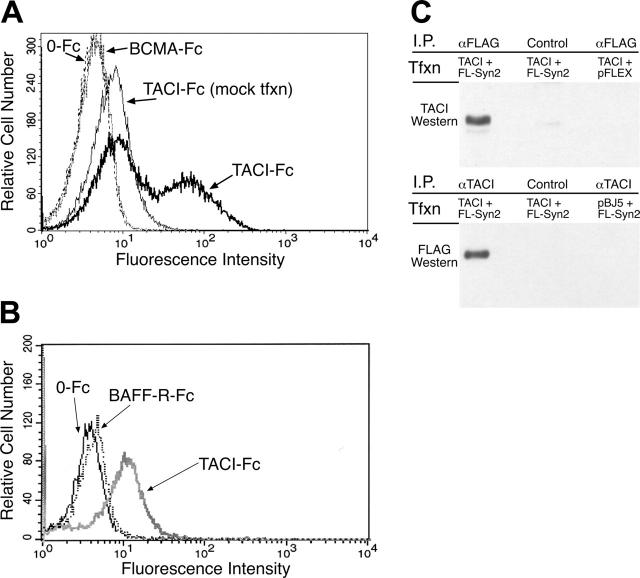
B-lymphocyte homeostasis and function are regulated by complementary actions of the TNFR family members TACI, BCMA, and BAFF-R, which are expressed by mature B cells. How these receptors are differentially activated is not entirely understood, because the primary ligand BAFF binds to all three. We searched for alternative ligands for TACI using recombinant TACI-Fc fusion protein as a probe and identified syndecan-2 as a new binding partner. TACI binding appears to require heparan sulfate posttranslational modifications of syndecan-2, because free heparin or pretreatment with heparitinase blocked the interaction. Syndecan-2 bound TACI but bound neither BAFF-R nor BCMA. Transfected cells expressing syndecan-2 activated signaling through TACI, as indicated by an NFAT-specific reporter. Syndecan-1 and syndecan-4 were also able to induce TACI signaling in a similar manner. This is the first identification of ligands that selectively activate TACI without simultaneously triggering BCMA or BAFF-R. This finding may help explain the alternative outcomes of signaling from this family of receptors in B cells.


Figures

Figure 1.
Figure 1.
TACI-Fc binds specifically to 293T cells. (A) Recombinant proteins containing (TACI-Fc; lanes 1 and 2) or lacking (0-Fc; lanes 3 and 4) the extracellular domain of TACI fused to the Fc region of human IgG1 were purified as described in “Materials and methods.” Proteins were analyzed by SDS-PAGE and detected by Coomassie blue staining. (B) Cells were examined for expression of putative TACI ligands by incubation with TACI-Fc, the control protein 0-Fc, or an antibody to the CD45 molecule as a positive control. Antibody and Fc binding were detected by flow cytometry. Numerous cell lines tested, including CEM cells, did not bind TACI-Fc, whereas (C) 293T cells showed significant binding to TACI-Fc but not to 0-Fc.
Figure 2.
Figure 2.
293T cells activate TACI signaling in a coculture assay. (A) To detect functional activation by a ligand on 293T cells, TAg Jurkat T cells were transiently transfected with an NFAT-SEAP reporter plasmid and a plasmid directing expression of full-length TACI (TACI), a truncated version lacking the cytoplasmic tail (TACI C-107), or empty vector (control). Equal aliquots of the transfected cells were treated as indicated, with PMA and ionomycin, providing an indication of the maximal activity of the NFAT reporter. For coculture, transfected TAg Jurkat cells were layered onto previously seeded and washed wells containing growing 293T or OV-1063 cells. NFAT-specific SEAP activity was determined and expressed as a fraction of PMA- and ionomycin-induced activity. 293T cells and TACI antibody induced significant activation of NFAT only in cells transfected with full-length TACI expression plasmid. (B) 293T cells do not express either BAFF or APRIL. mRNA was prepared from 293T cells and from known positive cell lines (HeLa for APRIL and HL-60 for BAFF), and RT-PCR was performed as described in “Materials and methods.” 293T cells do not contain detectable mRNA levels of either APRIL (left panel) or BAFF (right panel). (C) A 293T expression library was screened for binding partners with TACI-Fc and revealed syndecan-2 as a candidate. The extent of our syndecan-2 clone is shown here, where the region encoding the signal sequence is highlighted and the area coding for the transmembrane domain is underlined.
Figure 3.
Figure 3.
TACI-Fc binds to syndecan-2. (A-B) To verify that syndecan-2 was the interacting protein from the 293T library, TAg Jurkat cells were transiently transfected with an expression plasmid containing the syndecan-2 cDNA. Cells were stained with TACI-Fc, 0-Fc, BCMA-Fc (A), or BAFF-R-Fc (B), as indicated, and analyzed by flow cytometry. Some transfected cells bound to TACI-Fc, as shown by the right-shifted peak. Transient transfection does not deliver DNA to all cells in the culture; thus, some of the curve overlaps that of mock-transfected cells. (C) TACI coprecipitates with FLAG-tagged syndecan-2. 293T cells were transiently transfected with plasmids expressing TACI and FLAG epitope-tagged syndecan-2 (top and bottom panels, lanes 1 and 2), or TACI and the parent plasmid pFLEX (top, lane 3), or control plasmid pBJ5 and FLAG-tagged syndecan-2 (bottom, lane 3). Immunoprecipitations were carried out with antibodies specific for the FLAG epitope on syndecan-2 or for TACI, or control antibodies as indicated. Coprecipitated proteins were detected by Western blotting with TACI (top panel) or FLAG (bottom panel) antibodies.
Figure 4.
Figure 4.
TACI-Fc binding requires the heparan sulfate side chains of syndecan-2. (A) Parental Jurkat cells and stable Synd2/Jurkat cells were assayed for their ability to bind TACI-Fc in the presence or absence of heparin (as indicated). Addition of heparin completely blocked binding to syndecan-2-expressing Jurkat cells. (B) Treatment of Synd2/Jurkat cells with the heparan sulfate-cleaving enzymes heparinase or heparitinase reduced the ability of cells to bind TACI-Fc. Curves shown in panels A and B were analyzed by flow cytometry in the same experiment but are shown in separate panels for clarity. Negative and positive control curves (Jurkat and Synd2/Jurkat) in panels A and B are the same data. (C) Syndecan-2 does not bind a TACI ligand noncovalently. Synd2/Jurkats were incubated with heparin and then washed extensively to remove heparin and any putative ligands that might have been bound to cell surface syndecan-2. Washed Synd2/Jurkat cells recover their ability to bind TACI-Fc. (D) TACI-Fc binds directly to immobilized heparin or heparan sulfate. 0-Fc (lanes 3 and 4) or TACI-Fc (lanes 6 and 7) were mixed with carbozone resin conjugated to heparin (Hi) or heparan sulfate (Ha). Unbound fusion proteins were removed by extensive washing. Immobilized recombinant protein was subsequently eluted with SDS and analyzed by SDS-PAGE followed by Coomassie blue staining. Lanes 2 and 5 (labeled “S”) indicate the input amount of 0-Fc or TACI-Fc.
Figure 5.
Figure 5.
Syndecan-2 activates NFAT signaling through TACI. TACI-mediated activation of an NFAT-SEAP reporter plasmid was assayed. TAg Jurkat cells were transfected with a TACI expression plasmid together with NFAT reporter. These cells were cultured in the absence of any stimulation (no stimulation), control Jurkat cells, TACI antibody, or Synd2/Jurkat cells in the presence or absence of competing heparin as indicated. Experiments demonstrate that NFAT activation by syndecan-2-expressing cells is blocked by the competitor heparin. TACI antibody-mediated activation is, however, not blocked by heparin.
Figure 6.
Figure 6.
Syndecan-1 and syndecan-4 can also bind to and activate TACI. (A) TAg Jurkat cells were electroporated with either empty vector (broken line) or one of the indicated syndecan expression plasmids (solid line). Binding of TACI-Fc to syndecan-expressing cells was determined by flow cytometry. TACI-Fc binds to syndecans-1, -2, and -4 similarly. (B) TAg Jurkat cells were transiently transfected with empty vector plus NFAT reporter plasmid or a TACI cDNA with reporter plasmid, or with syndecan cDNAs. The TACI/NFAT cells were cocultured with either no stimulants, empty vector, or syndecan-expressing cells. Transfection of Jurkat cells with TACI/NFAT alone results in basal activation. Syndecan-1 and syndecan-4 activated NFAT-SEAP reporter similarly to syndecan-2. Expression of the syndecans in TAg Jurkat cells was verified by flow cytometry using heparan sulfate antibodies (bottom panel). (C) Heparin does not interfere with BAFF binding to TACI. TACI-Fc was incubated alone or with BAFF in the absence or presence of saturating levels of heparin (10 μg/mL). TACI-Fc was purified from solution by passage over protein A-Sepharose, and adsorbed BAFF eluted with SDS, and then detected by Western blotting. Recombinant BAFF migrates as 2 bands on SDS-PAGE, as indicated by arrows.
Figure 7.
Figure 7.
Syndecan-2 induces proliferation of B cells. (A) B220+ B cells from either wild-type (WT) or TACI knockout mice (KO) were cocultured with mitomycin C-treated Jurkat and Synd2/Jurkat cells at a ratio of 1:3 (105:3 × 10⁵). Coculture conditions included 10 ng/mL IL-4, 1 μg/mL anti-IgM F(ab′)2, 100 ng/mL BAFF, 1 μg/mL anti-CD40L, alone or in combination. Cells were incubated for 5 days, at which time cultures were pulsed with 1 μCi (0.037 MBq) 3H-TdR for 18 hours before harvesting. Stimulation of both WT and TACI-KO B cells with IL-4 and αIgM with or without αCD40L, together with either parental Jurkat or Synd2/Jurkat cells, resulted in minimal proliferation. With the addition of BAFF, B-cell growth conditions became more optimal, and a significant increase in proliferation of WT B cells was seen in response to Synd2/Jurkat. TACI-KO B cells also proliferated at a higher rate in response to Synd2/Jurkat cells but considerably less than did WT B cells. Parental Jurkat and Synd2/Jurkat cells showed minimal proliferation after mitomycin C treatment (far right). (B) CD19+ cells from human donors were cocultured with irradiated pEAK12/Jurkat or Synd2/Jurkat cells. Coculture was performed in the presence of 100 ng/mL BAFF, 2 μg/mL anti-IgM F(ab′)2, 100 ng/mL IL-4, alone or in combination. Cells were pulsed with tritiated thymidine as in Panel A. In the absence of stimulation, or presence of BAFF, αIgM, or BAFF plus αIgM, B-cell growth is minimal in response to both pEAK12/Jurkat and Synd2/Jurkat cells. In the presence of IL-4, BAFF plus IL-4, or αIgM plus IL-4, B-cell growth increased significantly, particularly in response to Synd2/Jurkat cells.

References

    1. Mackay F, Schneider P, Rennert P, Browning J. BAFF AND APRIL: a tutorial on B cell survival. Ann Rev Immunol. 2003;21: 231-264. - PubMed
    1. Bodmer JL, Schneider P, Tschopp J. The molecular architecture of the TNF superfamily. Trends Biochem Sci. 2002;27: 19-26. - PubMed
    1. Lentz VM, Cancro MP, Nashold FE, Hayes CE. Bcmd governs recruitment of new B cells into the stable peripheral B cell pool in the A/WySnJ mouse. J Immunol. 1996;157: 598-606. - PubMed
    1. Lentz VM, Hayes CE, Cancro MP. Bcmd decreases the life span of B-2 but not B-1 cells in A/WySnJ mice. J Immunol. 1998;160: 3743-3747. - PubMed
    1. Miller DJ, Hayes CE. Phenotypic and genetic characterization of a unique B lymphocyte deficiency in strain A/WySnJ mice. Eur J Immunol. 1991;21: 1123-1130. - PubMed

Publication types

MeSH terms

LinkOut - more resources