. 2006 Jan;116(1):137-45.
doi: 10.1172/JCI26121. Epub 2005 Dec 15.

A new type of radiosensitive T-B-NK+ severe combined immunodeficiency caused by a LIG4 mutation

Affiliations

A new type of radiosensitive T-B-NK+ severe combined immunodeficiency caused by a LIG4 mutation

Mirjam van der Burg et al. J Clin Invest. 2006 Jan.

Abstract

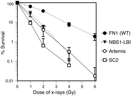
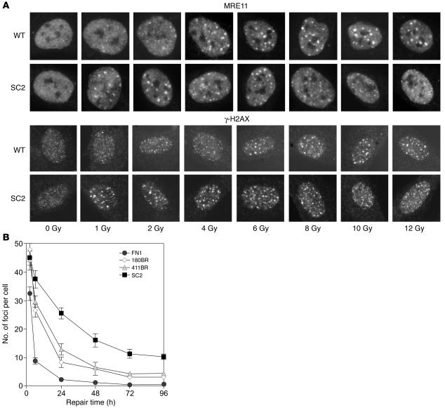
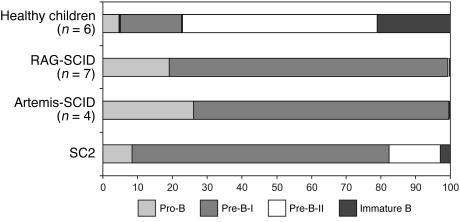
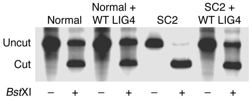
V(D)J recombination of Ig and TCR loci is a stepwise process during which site-specific DNA double-strand breaks (DSBs) are made by RAG1/RAG2, followed by DSB repair by nonhomologous end joining. Defects in V(D)J recombination result in SCID characterized by absence of mature B and T cells. A subset of T-B-NK+ SCID patients is sensitive to ionizing radiation, and the majority of these patients have mutations in Artemis. We present a patient with a new type of radiosensitive T-B-NK+ SCID with a defect in DNA ligase IV (LIG4). To date, LIG4 mutations have only been described in a radiosensitive leukemia patient and in 4 patients with a designated LIG4 syndrome, which is associated with chromosomal instability, pancytopenia, and developmental and growth delay. The patient described here shows that a LIG4 mutation can also cause T-B-NK+ SCID without developmental defects. The LIG4-deficient SCID patient had an incomplete but severe block in precursor B cell differentiation, resulting in extremely low levels of blood B cells. The residual D(H)-J(H) junctions showed extensive nucleotide deletions, apparently caused by prolonged exonuclease activity during the delayed D(H)-J(H) ligation process. In conclusion, different LIG4 mutations can result in either a developmental defect with minor immunological abnormalities or a SCID picture with normal development.


Figures

Figure 1
Figure 1
Clonogenic survival assay of fibroblasts after IR. Fibroblasts from patient SC2, the Artemis-deficient SCID patient (Artemis-1), and the NBS1-LBI cell line with an NBS1 mutation were radiosensitive. WT FN1 fibroblasts were used as normal control. Each survival curve represents the mean of at least 3 independent experiments. Error bars represent SEM.
Figure 2
Figure 2
IRIF formation in patient SC2 fibroblasts. (A) WT primary human fibroblasts and patient SC2 primary fibroblasts were untreated or γ irradiated with increasing doses (1, 2, 4, 6, 8, 10, and 12 Gy) and fixed after an incubation period of 8 hours followed by immunostaining with the MRE11 or γ-H2AX antibodies. (B) Kinetics of loss of γ-H2AX foci in fibroblasts derived from patient SC2 and the LIG4-defective patients 180BR (a radiosensitive leukemia patient) and 411BR (a patient with the LIG4 syndrome), and WT FN1 cells, following x-ray irradiation (1 Gy). The number of foci per nucleus was determined in 40 cells at the indicated times after irradiation.
Figure 3
Figure 3
Flow cytometric analysis of BM precursor B cell compartment. Composition of the BM precursor B cell compartment in the patient, healthy controls (n = 6), RAG-deficient SCID patients (n = 7), Artemis-deficient patients (n = 4), and patient SC2. RAG-deficient and Artemis-deficient SCID patients showed a complete block before the CyIgμ-positive pre-B-II cell stage, whereas patient SC2 showed an incomplete block, with approximately 20% pre-B-II and immature B cells.
Figure 4
Figure 4
DNA end joining. (A) A linear substrate with homologous ends (ATCAGC) can be rejoined via direct end joining or via microhomology-directed end joining. Joining via microhomology results in the generation of a BstXI restriction site (CCAN6TGG). (B) Analysis of joining products in WT (Normal), SC2, Artemis (Artemis-1), and LIG4 mutant (180BR) cells. Junctions were PCR amplified, and PCR products were digested with BstXI, as indicated.
Figure 5
Figure 5
Analysis of LIG4 protein expression. (A) Western blot analysis of LIG4 protein expression showed absence of an approximately 100-kDa band in patient SC2. Artemis-1 was used as positive control. The position of the 100-kDa band of the molecular weight marker is indicated on the left. (B) After immunoprecipitation of the LIG4/XRCC4 complex with the XRCC4 antibody, no LIG4 protein was detectable in patient SC2.
Figure 6
Figure 6
Complementation of end-joining defect with the WT LIG4. Analysis of joining products of the linear substrate with homologous ends (Figure 4A) in WT and SC2 patients with and without cotransfection of a WT LIG4 expression construct. Junctions were PCR amplified, and PCR products were digested with BstXI, as indicated.

References

    1. Schatz DG. V(D)J recombination. Immunol. Rev. 2004;200:5–11. - PubMed
    1. Gellert M. V(D)J recombination: RAG proteins, repair factors, and regulation. Annu. Rev. Biochem. 2002;71:101–132. - PubMed
    1. McBlane JF, et al. Cleavage at a V(D)J recombination signal requires only RAG1 and RAG2 proteins and occurs in two steps. Cell. 1995;83:387–395. - PubMed
    1. Van Gent DC, Ramsden DA, Gellert M. The RAG1 and RAG2 proteins establish the 12/23 rule in V(D)J recombination. Cell. 1996;85:107–113. - PubMed
    1. Lees-Miller SP, Meek K. Repair of DNA double strand breaks by non-homologous end joining. Biochimie. 2003;85:1161–1173. - PubMed

Publication types