. 2005;12(4-6):289-303.
doi: 10.3727/000000005783992034.

A selective role for MRF4 in innervated adult skeletal muscle: Na(V) 1.4 Na+ channel expression is reduced in MRF4-null mice

Affiliations

A selective role for MRF4 in innervated adult skeletal muscle: Na(V) 1.4 Na+ channel expression is reduced in MRF4-null mice

Amy L Thompson et al. Gene Expr. 2005.

Abstract

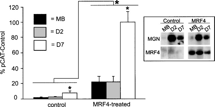
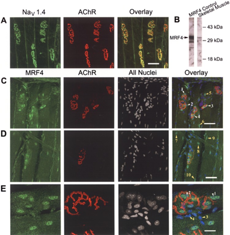
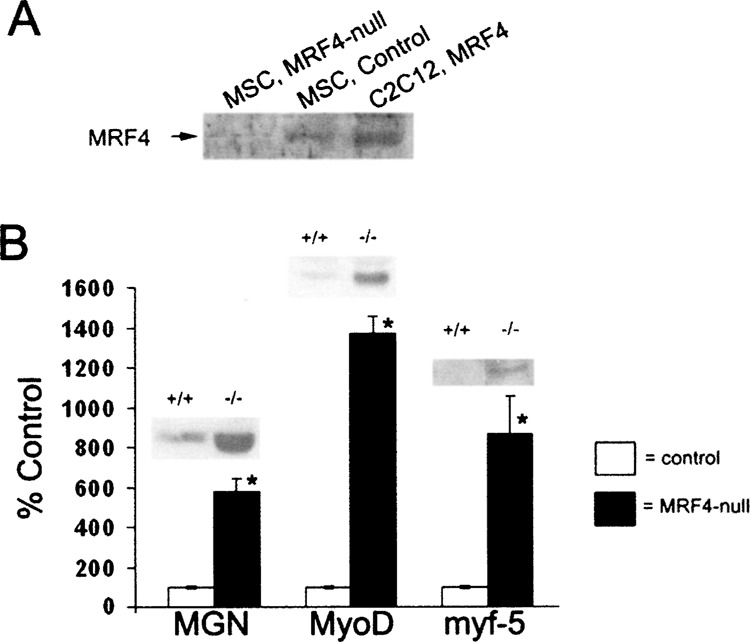
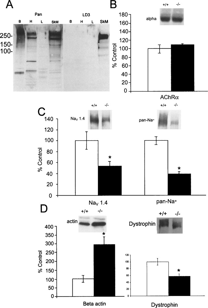
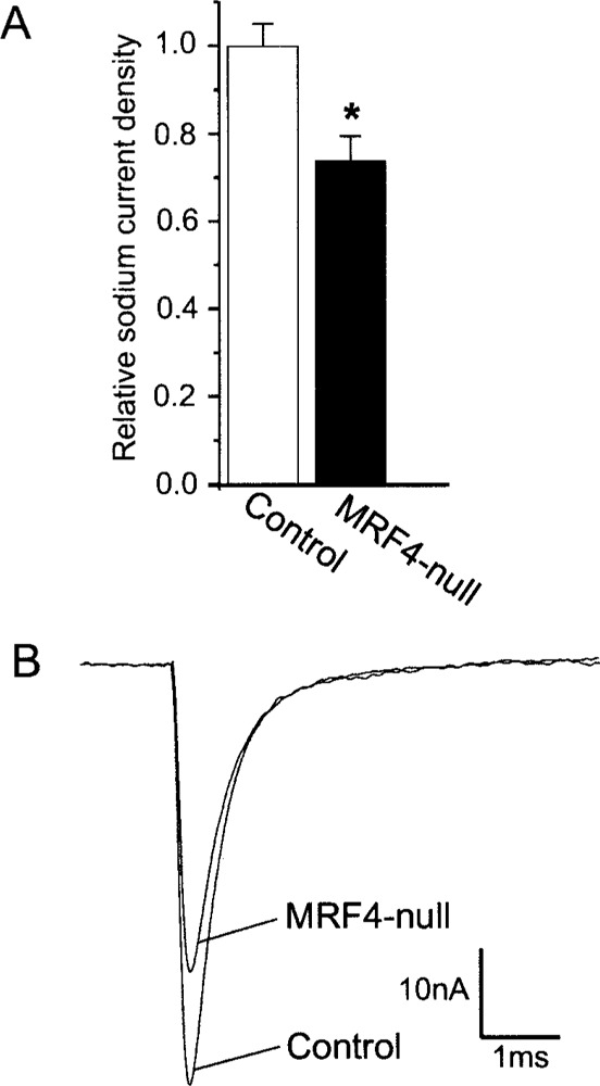
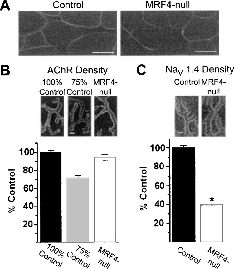
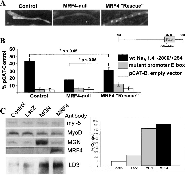
The factors that regulate transcription and spatial expression of the adult skeletal muscle Na+ channel, Na(V) 1.4, are poorly understood. Here we tested the role of the transcription factor MRF4, one of four basic helix-loop-helix (bHLH) factors expressed in skeletal muscle, in regulation of the Na(V) 1.4 Na+ channel. Overexpression of MRF4 in C2C12 muscle cells dramatically elevated Na(V) 1.4 reporter gene expression, indicating that MRF4 is more efficacious than the other bHLH factors expressed at high levels endogenously in these cells. In vivo, MRF4 protein was found both in extrajunctional and subsynaptic muscle nuclei. To test the importance of MRF4 in Na(V) 1.4 gene regulation in vivo, we examined Na+ channel expression in MRF4-null mice using several techniques, including Western blotting, immunocytochemistry, and electrophysiological recording. By all methods, we found that expression of the Na(V) 1.4 Na+ channel was substantially reduced in MRF4-null mice, both in the surface membrane and at neuromuscular junctions. In contrast, expression of the acetylcholine receptor, and in particular its alpha subunit, was unchanged, indicating that MRF4 regulation of Na+ channel expression was selective. Expression of the bHLH factors myf-5, MyoD, and myogenin was increased in MRF4-null mice, but these factors were not able to fully maintain Na(V) 1.4 Na+ channel expression either in the extrajunctional membrane or at the synapse. Thus, MRF4 appears to play a novel and selective role in adult muscle.


Figures

Figure 1
Figure 1
MRF4 increases NaV 1.4 reporter gene expression in C2C12 muscle cells. The −2800/+254 NaV 1.4 CAT reporter gene was transfected into C2C12 myoblasts along with the empty pCI expression vector (control) or a MRF4 expression vector driven by the EMSV promoter (MRF4 treated). Cells were harvested as myoblasts (MB), day 2 myotubes (D2), or day 7 myotubes (D7). Reporter gene expression was assessed relative to that for a control plasmid, pCAT-Control. Averages are shown with error bars indicating SEM (n = 4). MRF4 increased NaV 1.4 reporter gene expression, especially in day 7 myotubes. Inset: Western blot analysis was carried out with antibodies to myogenin or MRF4 to monitor expression of endogenous and exogenous bHLH factors.
Figure 2
Figure 2
NaV 1.4 Na+ channels and MRF4 are expressed robustly in synaptic region. The sternomastoid muscle was fixed and processed for confocal microscopy as outlined in Materials and Methods. En face views of the synaptic region are shown in each panel. (A) NaV 1.4 Na+ channels (green) are expressed throughout the surface membrane, but are concentrated at neuromuscular junctions (NMJs) labeled with αBTX, which binds the synaptic marker, acetylcholine receptors (AChRs, red). Scale bar: 50 μm for (A, C, D). (B) Western blot analysis of a nuclear protein fraction demonstrated that the MRF4 antibody is specific for MRF4 in adult skeletal muscle. An MRF4 protein control was produced in cell culture using an expression vector for MRF4. (C, D) Nuclei associated with NMJs (AChRs, red) express MRF4 robustly (green nuclei indicated by white arrows 1–4). There are also MRF4-expressing nuclei in extrajunctional areas (green nuclei indicated by arrows 5–10). The unstained nuclei in the region of NMJs are likely those of the overlaying Schwann cells, which are seen more clearly in (E). (E) A close-up of a single NMJ. Scale bar: 20 μm. There is clear MRF4 staining of subsynaptic nuclei (white arrows 1 and 2). Additionally, there are unlabeled nuclei of a different morphology that lie predominantly outside of the myofiber (yellow arrows 3 and 4). Based on both morphology and location, these nuclei are likely those of the overlaying Schwann cells.
Figure 3
Figure 3
Other bHLH factors are expressed at higher levels in MRF4-null mice. (A) A Western blot with an MRF4 antibody was carried out on nuclear extracts prepared from muscle satellite cells derived from MRF4-null or control mice. A MRF4 expression vector was introduced into C2C12 cells to produce a positive control. (B) Western blot analysis of myogenin, MyoD, and myf-5 protein expression was carried out for both control and MRF4-null animals. Consistent with previously published results, expression of myogenin is increased in MRF4-null mice (18), and, for the first time, MyoD and myf-5 proteins are also found to be increased. Possibly these factors compensate for the absence of MRF4 in the regulation of many muscle genes.
Figure 4
Figure 4
Expression of AChR α subunit is unchanged in MRF4-null mice, but expression of NaV 1.4 Na+ channel is reduced. (A) Western blot analysis of LD3 immunoreactivity demonstrates that this antibody detects only the NaV 1.4 isoform expressed in adult skeletal muscle and not other sodium channel isoforms, including the NaV 1.5 isoform expressed in cardiac muscle, whereas the pan-Na+ channel antibody, to the highly conserved III-IV linker (Upstate Biotech), recognizes all isoforms. Labels indicate brain (Br), heart (H), liver (L), and adult skeletal muscle (SkM). The liver serves as a negative control. (B) Western blot analyses of the α subunit of the acetylcholine receptor (AChR) indicated that there is not a significant change in this protein. (C) Western blot analyses of Na+ channel were carried out using either the LD3 antibody specific for the NaV 1.4 Na+ channel (24) or a pan-Na+ channel antibody. Global expression of Na+ channels is reduced by approximately 50% (*p < 0.05, Student’s t-test). Because similar results were observed with both antibodies, these data indicate that expression of the adult NaV 1.4 Na+ channel is reduced, but there is no compensatory increase in expression of the embryonic NaV 1.5 Na+ channel. (D) Western blot analysis of β-actin expression was initially carried out with the goal of normalizing expression of other proteins to it, as is routinely done in many studies. However, we observed that expression of β-actin is increased in the MRF4-null animals (*p < 0.05), precluding its use in this manner. Additionally, dystrophin was found to be decreased in the absence of MRF4. Western blots were quantified by imaging on a Molecular Dynamics Storm PhosphorImager and samples normalized to the average of the control samples (n = 5 control animals and 7 MRF4-null animals).
Figure 5
Figure 5
Peak Na+ channel current density is reduced in the extrajunctional surface membrane of MRF4-null mice. (A) A plot of the average relative Na+ density is shown (*p < 0.01, n = 9 pairs of MRF4-null and control animals). Currents were measured using the technique of loose-patch clamp as described previously (36) and in Materials and Methods. (B) The peak inward Na+ currents observed for representative muscle fibers from an MRF4-null and control mouse pair are superimposed.
Figure 6
Figure 6
Expression of NaV 1.4 Na+ channels, but not AChRs, is reduced at neuromuscular junctions in MRF4-null mice. (A) Muscle cross sections stained with a directly conjugated NaV 1.4-specific antibody in both control and MRF4-null muscle. Na+ channels are inserted into the surface membrane normally, although there appears to be a reduction in intensity in the MRF4-null mice relative to the control. Scale bar: 30 μm. (B) Analysis of en face views of NMJs stained with rhodamine-αBTX in both control and MRF4-null animals. Despite our ability to detect differences in fluorescence to within 5%, we could detect no reduction in AChR density at NMJs from MRF4-null mice. (C) Analysis of en face views of NMJs stained with directly conjugated NaV 1.4-specific antibody. There is a 60% reduction in Na+ channel density at neuromuscular junctions in MRF4-null animals (*p < 0.01, n = 6 animals). Scale bar in (B, C): 2 μm.
Figure 7
Figure 7
MRF4 regulates NaV 1.4 gene expression at the transcriptional level. Either a wild-type or the promoter E box mutant NaV 1.4 reporter gene was introduced into muscle satellite cell cultures, derived from either control or MRF4-null animals (B). Expression was normalized relative to that of a control vector, pCAT-Control. As a negative control, an empty expression vector, pCAT-B, was also used. Expression of the wild-type reporter gene is reduced in MRF4-null cultures, but is partially “rescued” by treating these cultures with 100 MOI of a MRF4 adenovirus. The promoter E box mutation decreases expression to nearly background levels under all conditions examined. Sister cultures of those used for transfections were analyzed for MRF4 protein by immunocytochemistry. As expected, MRF4 is expressed only in control and MRF4-rescued cultures, although the cellular distribution of MRF4 is both cytoplasmic and nuclear in the control cultures (A). To determine which bHLH factor might be compensating in the absence of MRF4, C2C12 myoblasts expressing only myf-5 and MyoD were infected with myogenin and MRF4 adenovirus (C). LacZ adenovirus was used as a negative control. Whereas myf-5 and MyoD have little effect on Na+ channel expression, myogenin can almost fully compensate.

Similar articles

Cited by

References

    1. Abbott K. L.; Friday B. B.; Thaloor D.; Murphy T. J.; Pavlath G. K. Activation and cellular localization of the cyclosporine A-sensitive transcription factor NF-AT in skeletal muscle cells. Mol. Biol. Cell 9:2905–2916; 1998. - PMC - PubMed
    1. Arber S.; Burden S. J.; Harris A. J. Patterning of skeletal muscle. Curr. Opin. Neurobiol. 12:100–103; 2002. - PubMed
    1. Arnold H. H.; Braun T. Genetics of muscle determination and development. Curr. Top. Dev. Biol. 48:129–164; 2000. - PubMed
    1. Awad S. S.; Lightowlers R. N.; Young C.; Chrzanowska-Lightowlers Z. M.; Lomo T.; Slater C. R. Sodium channel mRNAs at the neuromuscular junction: Distinct patterns of accumulation and effects of muscle activity. J. Neurosci. 21:8456–8463; 2001. - PMC - PubMed
    1. Bailey S. J.; Stocksley M. A.; Buckel A.; Young C.; Slater C. R. Voltage-gated sodium channels and ankyrinG occupy a different postsynaptic domain from acetylcholine receptors from an early stage of neuromuscular junction maturation in rats. J. Neurosci. 23:2102–2111; 2003. - PMC - PubMed

Publication types

MeSH terms

Substances

LinkOut - more resources