. 2006;138(1):17-24.
doi: 10.1016/j.neuroscience.2005.11.002. Epub 2005 Dec 15.

Glutamate-induced Ca2+ influx in third-order neurons of salamander retina is regulated by the actin cytoskeleton

Affiliations

Glutamate-induced Ca2+ influx in third-order neurons of salamander retina is regulated by the actin cytoskeleton

A Akopian et al. Neuroscience. 2006.

Abstract

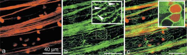
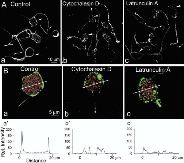
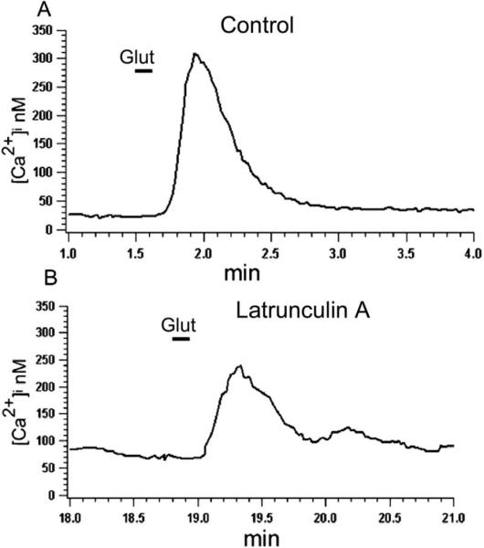
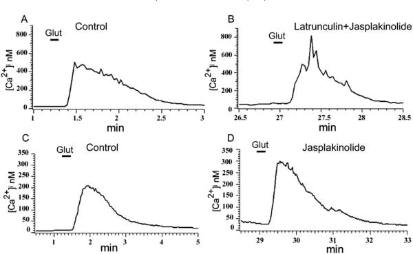
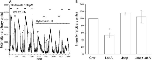
Ligand-gated ion channels (ionotropic receptors) link to the cortical cytoskeleton via specialized scaffold proteins and thereby to appropriate signal transduction pathways in the cell. We studied the role of filamentous actin in the regulation of Ca influx through glutamate receptor-activated channels in third-order neurons of salamander retina. Staining by Alexa-Fluor 488-phalloidin, to visualize polymerized actin, we show localization of filamentous actin in neurites, and the membrane surrounding the cell soma. With Ca(2+) imaging we found that in dissociated neurons, depolymerization of filamentous actin by latrunculin A, or cytochalasin D significantly reduced glutamate-induced intracellular Ca(2+) accumulation to 53+/-7% of control value. Jasplakinolide, a stabilizer of filamentous actin, by itself slightly increased the glutamate-induced Ca(2+) signal and completely attenuated the inhibitory effect when applied in combination with actin depolymerizing agents. These results indicate that in salamander retinal neurons the actin cytoskeleton regulates Ca(2+) influx through ionotropic glutamate receptor-activated channels, suggesting regulatory roles for filamentous actin in a number of Ca(2+)-dependent physiological and pathological processes.


Figures

Fig. 1
Fig. 1
Distribution of F-actin in retinal ganglion cells. Ganglion cells and axon fibers were retrogradely labeled with rhodamine-dextran (A), and then flat-mounts stained with Alexa Fluor 488-phalloidin (B). F-actin was present in axonal fibers and numerous processes of ganglion cells; often appearing as bright spots (inset, arrowheads). Merger of two confocal images is illustrated in C. The high-power view of ganglion cells (inset) was obtained by focusing on cell soma labeled with dextran (red), and phalloidin (green).
Fig. 2
Fig. 2
Disruption of the actin cytoskeleton in retinal neurons by latrunculin A. (A) Confocal images of dissociated third-order retinal neurons exposed for 30 min to either vehicle (Control), or 10 μM cytochalasin D, or 5 μM latrunculin A before fixation and staining with Alexa-Fluor488-phalloidin. Note intense fluorescence ring surrounding the cell perimeter (arrowheads) in control cells, and predominantly punctate character of staining, or loss of fluorescence in cytochalasin-, and latrunculin-treated cells, respectively. (B) F-actin staining in dissociated rhodamine-dextran-labeled ganglion cells in control saline (a), and after incubation with cytochalasin D (b), or latrunculin A (c). (a′–c′) Shows the phalloidin fluorescence intensity profile across the white line marked in a–c.
Fig. 3
Fig. 3
Effect of latrunculin A on the glutamate-induced Ca2+ influx. In control solution glutamate (100 μM) induced Ca2+ influx (A), which was significantly (P<0.05) reduced following 30 min incubation of cells with 5 μM latrunculin A (B).
Fig. 4
Fig. 4
Jasplakinolide attenuates the inhibitory effect of latrunculin A on glutamate-induced Ca2+ influx. Cells were incubated for 30 min in Ringer solution containing 5 μM latrunculin A+5 μM jasplakinolide (B), or jasplakinolide alone (D), and glutamate-induced Ca2+ signals were compared with those measured in control solution (A and C). Neither jasplakinolide alone nor in combination with latrunculin A had statistically significant effect on the glutamate-induced Ca2+ signal (P>0.3 and P>0.1 respectively).
Fig. 5
Fig. 5
Effect of cytochalasin D on Ca2+ influx via glutamate receptor-activated channels, and voltage-activated Ca2+ channels. (A) High-temporal resolution Ca2+ signals induced by glutamate (300 μM), and KCl (20 mM) were recorded with a confocal microscope before and after incubation of cells with 10 μM cytochalasin D. Cytochalasin D itself caused a transient increase in [Ca2+]i (arrowhead), and reduced both glutamate-, and KCl-induced Ca2+ signals. The amplitude of spontaneous Ca2+ transients (asterisks) was also reduced following exposure to cytochalasin D. (B) Bar graph summarizing the effect of F-actin depolymerization on glutamate-induced Ca2+ accumulation.

Similar articles

Cited by

References

    1. Akopian A, Witkovsky P. Calcium and retinal function. Mol Neurobiol. 2002;2:113–132. - PubMed
    1. Berdiev BK, Prat AG, Centiello HF, Ausiello DA, Fuller CM, Jovoy B, Benos DJ, Ismailov II. Regulation of epithelial sodium channels by short actin filaments. J Biol Chem. 1996;271:17704–17710. - PubMed
    1. Berridge MJ, Bootman MD, Roderick HL. Calcium signalling: dynamics, homeostasis and remodeling. Nat Rev Mol Cell Biol. 2003;4:517–529. - PubMed
    1. Bubb MR, Senderowicz AM, Sausville EA, Duncan KL, Korn ED. Jasplakinolide, a cytotoxic natural product, induces actin polymerization and competitively inhibits the binding of phalloidin to F-actin. J Biol Chem. 1994;269:14869–14871. - PubMed
    1. Carroll RC, Beattie EC, Xia H, Luscher C, Altschuler Y, Nicoll RA, Malenka RC, von Zastrow M. Dynamin-dependent endocytosis of ionotropic glutamate receptors. Proc Natl Acad Sci U S A. 1999;96:14112–14117. - PMC - PubMed

Publication types

MeSH terms

LinkOut - more resources