. 2005 Dec 16;123(6):1051-63.
doi: 10.1016/j.cell.2005.09.035.

HIM-8 binds to the X chromosome pairing center and mediates chromosome-specific meiotic synapsis

Affiliations

HIM-8 binds to the X chromosome pairing center and mediates chromosome-specific meiotic synapsis

Carolyn M Phillips et al. Cell. .

Abstract

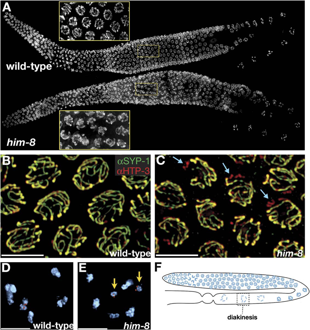
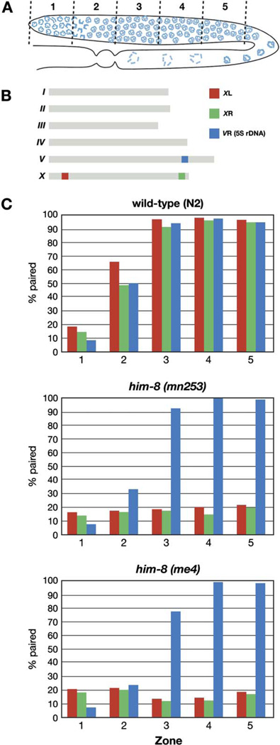
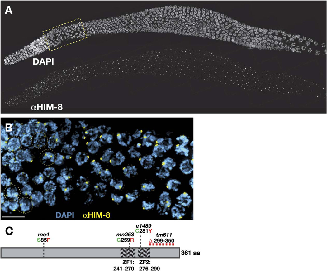
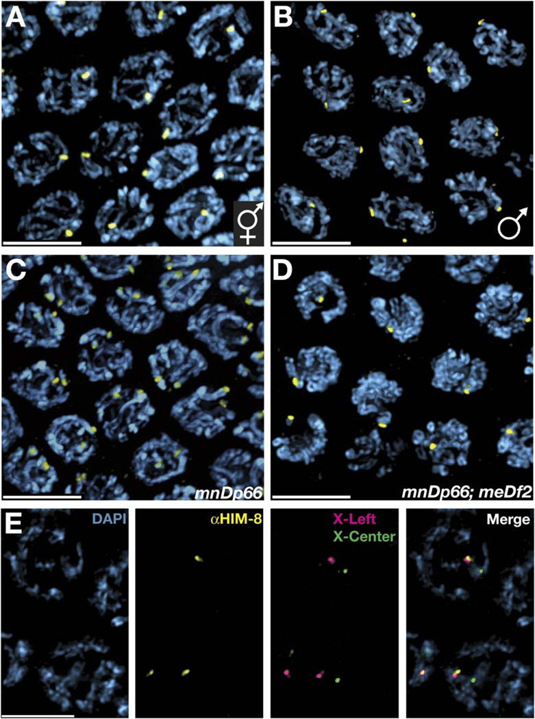
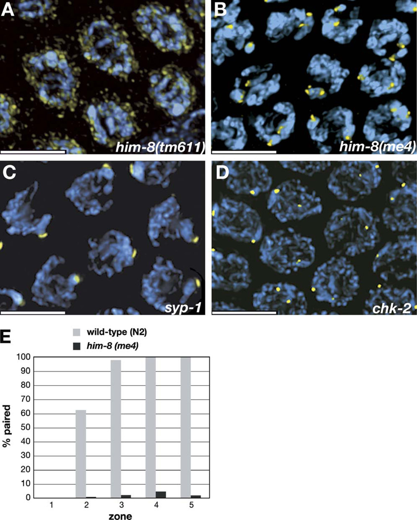
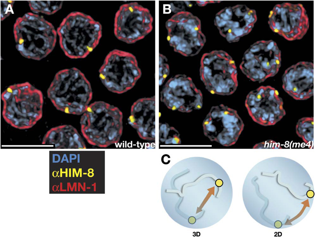
The him-8 gene is essential for proper meiotic segregation of the X chromosomes in C. elegans. Here we show that loss of him-8 function causes profound X chromosome-specific defects in homolog pairing and synapsis. him-8 encodes a C2H2 zinc-finger protein that is expressed during meiosis and concentrates at a site on the X chromosome known as the meiotic pairing center (PC). A role for HIM-8 in PC function is supported by genetic interactions between PC lesions and him-8 mutations. HIM-8 bound chromosome sites associate with the nuclear envelope (NE) throughout meiotic prophase. Surprisingly, a point mutation in him-8 that retains both chromosome binding and NE localization fails to stabilize pairing or promote synapsis. These observations indicate that stabilization of homolog pairing is an active process in which the tethering of chromosome sites to the NE may be necessary but is not sufficient.


Figures

Figure 1
Figure 1. him-8 Mutants Display X Chromosome-Specific Defects in Synapsis and Chiasma Formation
(A) Whole gonads from wild-type and him-8 hermaphrodites stained with DAPI. Insets magnify the pachytene region to show that him-8 nuclei retain a polarized appearance that is usually restricted to transition-zone nuclei. (B and C) Pachytene nuclei stained with antibodies to SYP-1 (green) and HTP-3 (red). (B) In wild-type hermaphrodites, these antibodies show very similar localization patterns at pachytene along the entire lengths of all six pairs of chromosomes. (C) In him-8 mutant hermaphrodites we observe one pair of unsynapsed chromosomes in each nucleus. Here these are revealed as chromosomes that stain with HTP-3 (red) but not SYP-1 (green). Examples are indicated with blue arrows. (D and E) Oocyte nuclei at diakinesis, shortly prior to the meiotic nuclear divisions. (D) Wild-type nuclei have six DAPI staining bodies, indicating the formation of chiasmata between all six pairs of homologous chromosomes. One of these bivalents is marked by a FISH probe specific for the X chromosomes. (E) him-8 nuclei usually reveal seven DAPI staining bodies at diakinesis. Achiasmate, or univalent, X chromosomes marked by a FISH probe are indicated by yellow arrows. (F) Diagram showing the location of diakinesis within the worm gonad. At this stage, the SC has largely broken down and homologs are held together by chiasmata. All images are projections of 3D images following deconvolution. Scale bars represent 5 µm.
Figure 2
Figure 2. X Chromosome Pairing Is Defective in him-8 Mutants
(A) Diagram of a hermaphrodite gonad, indicating the five zones in which the pairing of FISH signals was scored. (B) Genomic localizations of the three FISH probes used to quantify homolog pairing. (C) Graphs indicating the fraction of paired FISH signals in each zone for wild-type (N2), him-8(mn353), and him-8(me4) hermaphrodites. Three probes were scored independently: one from the left end of X chromosome (red), one from the right end of X chromosome (green), and the 5S rDNA, which marks the right arm of chromosome V (blue). In both him-8 alleles, pairing of the X chromosome probes did not rise above the baseline levels observed in the premeiotic region (zone 1), whereas Chromosome V association rates and dynamics were very similar to what we observed in wild-type hermaphrodites.
Figure 3
Figure 3. HIM-8 is a C2H2 Zinc-Finger Protein that Localizes to Distinct Nuclear Foci during Meiosis
(A) Three-dimensional projection through a wild-type gonad stained with DAPI and antibodies against HIM-8. Subnuclear HIM-8 foci are present in all germ-line nuclei throughout the premeiotic, transition-zone, and pachytene region of the gonad. The transition-zone region outlined by the yellow box is magnified in (B). (B) Prior to meiotic entry, two HIM-8 foci (yellow) are observed in each nucleus. Examples of premeiotic nuclei in which two foci can be clearly observed are outlined in brown circles. Once nuclei have entered the transition zone, representing the leptotene/zygotene stages of meiosis, they usually reveal only a single HIM-8 focus or closely spaced pair of foci. The scale bar represents 5 µm. (C) Schematic representation of the HIM-8 protein. The diagram displays the location of two predicted zinc fingers as well as the sites of point mutations or deletions resulting from the four mutant alleles of him-8.
Figure 4
Figure 4. The HIM-8 Protein Localizes to the PC Region of the X Chromosome
All images show projections through fields of pachytene-region nuclei from animals of the indicated genotypes. The entire nuclear volume is shown in each projection. All scale bars represent 5 µm. (A) Wild-type hermaphrodite, revealing a single HIM-8 focus in each nucleus due to close association between the two binding sites detected in premeiotic and very early meiotic nuclei (Figure 3). (B) Wild-type male, which displays a single (unpaired) HIM-8 focus in each nucleus. In contrast to hermaphrodites, males also have one HIM-8 focus per nucleus in the premeiotic region of the gonad (data not shown). (C) Hermaphrodite carrying two copies of the mnDp66 duplication of the X chromosome PC region. An additional HIM-8 focus is present in each nucleus, corresponding to the paired and synapsed duplication. (D) Hermaphrodite carrying both mnDp66 and the PC deficiency meDf2, which eliminates one of the two foci observed in (C). (E) HIM-8 immunostaining was performed in conjunction with FISH to two probes on the X chromosome, one from the left arm (1.5 Mb from the telomere) and one from a more medial position (7.4 Mb from the left end). The XL probe is closely associated with HIM-8 focus, while the XC probe is clearly more distant from the HIM-8 focus. Probes to the right end of the X and autosomal loci were also tested (data not shown) and did not correlate in their position with HIM-8 foci.
Figure 5
Figure 5. HIM-8 Localization in him-8 Mutants and Other Informative Meiotic Mutants
All images show projections through the nuclear volumes of fields of pachytene-region nuclei from animals of the indicated genotypes. Immunofluorescence with anti-HIM-8 antibodies is shown in yellow, and DAPI staining is shown in blue. (A) In him-8(tm611) mutant hermaphrodites, discrete foci of HIM-8 are not detected on the chromosomes. The intensity of the staining is shown more brightly here than in images of other genotypes to reveal that the residual staining is concentrated at the nuclear periphery. Similar staining is seen in him-8(e1489) and him-8(mn253) animals, which also carry mutations in the zinc-finger domain of HIM-8. This residual staining is detected using several different antisera raised against HIM-8, suggesting that it is specific rather than nonspecific background. (B) In him-8(me4) hermaphrodites, two distinct foci are visible in each nucleus at pachytene. See also Figure 6B and Figure S1. (C) In syp-1 hermaphrodites, a single focus of HIM-8 is detected in each nucleus in the pachytene region of the gonad, indicating that pairing of the HIM-8 binding region is stabilized despite the absence of synapsis. (D) HIM-8 foci are detected on the X chromosomes but usually do not pair in chk-2 mutants. (E) Immunofluorescence with the HIM-8 antibody was performed on wild-type and him-8(me4) hermaphrodites. The fraction of paired foci was scored in each of five zones of the gonad, which were defined in the same way as in our FISH analysis (Figure 2). All scale bars represent 5 µm.
Figure 6
Figure 6. HIM-8 Foci Associate with Nuclear-Envelope Components in Both Wild-Type and him-8(me4) Animals
(A and B) Pachytene nuclei were stained with antibodies against HIM-8 (yellow) and anti-LMN-1 (red), which marks the nuclear lamina, or nuclear envelope. All scale bars represent 5 µm. See Figure S1 for an optical section through a similar data set, where nuclear-envelope association is somewhat more evident. (C) A possible role for nuclear-envelope association of meiotic chromosomes is the reduction of homology search from a 3D to a 2D problem. As discussed in the text, our analysis of the him-8(me4) mutant indicates that the requirement for HIM-8 is likely to extend beyond such a role.

Comment in

Similar articles

Cited by

References

    1. Alpi A, Pasierbek P, Gartner A, Loidl J. Genetic and cytological characterization of the recombination protein RAD-51 in Caenorhabditis elegans . Chromosoma. 2003;112:6–16. - PubMed
    1. Anuradha S, Muniyappa K. Meiosis-specific yeast Hop1 protein promotes synapsis of double-stranded DNA helices via the formation of guanine quartets. Nucleic Acids Res. 2004a;32:2378–2385. - PMC - PubMed
    1. Anuradha S, Muniyappa K. Saccharomyces cerevisiae Hop1 zinc finger motif is the minimal region required for its function in vitro. J. Biol. Chem. 2004b;279:28961–28969. - PubMed
    1. Baker BS, Carpenter AT. Genetic analysis of sex chromosomal meiotic mutants in Drosophilia melanogaster . Genetics. 1972;71:255–286. - PMC - PubMed
    1. Bhalla N, Dernburg AF. A conserved checkpoint monitors meiotic chromosome synapsis in Caenorhabditis elegans . Science. 2005;310 in press. - PubMed

Publication types

MeSH terms