

Molecular pathways mediating mechanical signaling in bone
- PMID: 16361069
- PMCID: PMC3687520
- DOI: 10.1016/j.gene.2005.10.028
Molecular pathways mediating mechanical signaling in bone
Abstract
Bone tissue has the capacity to adapt to its functional environment such that its morphology is "optimized" for the mechanical demand. The adaptive nature of the skeleton poses an interesting set of biological questions (e.g., how does bone sense mechanical signals, what cells are the sensing system, what are the mechanical signals that drive the system, what receptors are responsible for transducing the mechanical signal, what are the molecular responses to the mechanical stimuli). Studies of the characteristics of the mechanical environment at the cellular level, the forces that bone cells recognize, and the integrated cellular responses are providing new information at an accelerating speed. This review first considers the mechanical factors that are generated by loading in the skeleton, including strain, stress and pressure. Mechanosensitive cells placed to recognize these forces in the skeleton, osteoblasts, osteoclasts, osteocytes and cells of the vasculature are reviewed. The identity of the mechanoreceptor(s) is approached, with consideration of ion channels, integrins, connexins, the lipid membrane including caveolar and non-caveolar lipid rafts and the possibility that altering cell shape at the membrane or cytoskeleton alters integral signaling protein associations. The distal intracellular signaling systems on-line after the mechanoreceptor is activated are reviewed, including those emanating from G-proteins (e.g., intracellular calcium shifts), MAPKs, and nitric oxide. The ability to harness mechanical signals to improve bone health through devices and exercise is broached. Increased appreciation of the importance of the mechanical environment in regulating and determining the structural efficacy of the skeleton makes this an exciting time for further exploration of this area.
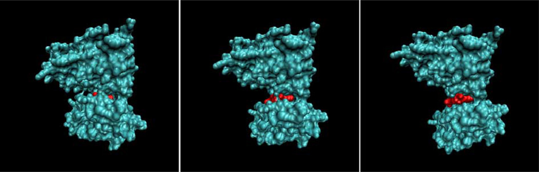
Figures


References
-
- Aikawa R, et al. Integrins play a critical role in mechanical stress-induced p38 MAPK activation. Hypertension. 2002;39:233–238. - PubMed
-
- Alford AI, Jacobs CR, Donahue HJ. Oscillating fluid flow regulates gap junction communication in osteocytic MLO-Y4 cells by an ERK1/2 MAP kinase-dependent mechanism. Bone. 2003;33:64–70. - PubMed
-
- Anderson RG. The caveolae membrane system. Annu. Rev. Biochem. 1998;67:199–225. - PubMed
-
- Armour KE, et al. Defective bone formation and anabolic response to exogenous estrogen in mice with targeted disruption of endothelial nitric oxide synthase. Endocrinology. 2001;142:760–766. - PubMed
-
- Baldwin KM, et al. Musculoskeletal adaptations to weightlessness and development of effective countermeasures. Med. Sci. Sports Exerc. 1996;28:1247–1253. - PubMed
Publication types
MeSH terms
Substances
Grants and funding
LinkOut - more resources
Full Text Sources
Other Literature Sources
