

Virtual colonoscopy for colorectal cancer screening: current status
- PMID: 16361129
- PMCID: PMC1665314
- DOI: 10.1102/1470-7330.2005.0108
Virtual colonoscopy for colorectal cancer screening: current status
Abstract
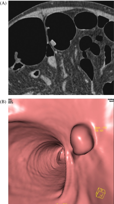
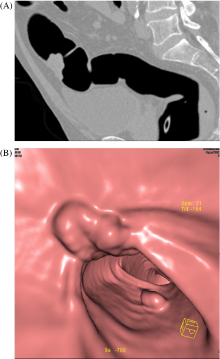
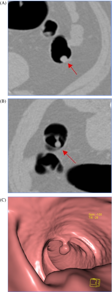
Computed tomography colonography (CTC) (also known as 'virtual colonoscopy') is a noninvasive method of imaging the colon using helical CT. Although CTC has been shown to be useful for certain clinical indications, it has not yet been endorsed as a colorectal cancer screening test. The purpose of this article is to review the current status of CTC for colorectal cancer screening. CTC is an accurate method to detect colonic polyps and to select patients who would benefit from colonoscopy. The major advantages of CTC over conventional colonography include its relatively low risk and greater tolerance by patients. In this article, the CTC procedure and results of clinical trials are reviewed, as well as potential pitfalls related to CTC performance and interpretation. Finally, radiation dose, the discovery of incidental extracolonic findings with CTC, bowel preparation methods, and computer-aided diagnosis are addressed.
International Cancer Imaging Society.
Figures
Similar articles
-
Current role and future potential of computed tomographic colonography for colorectal polyp detection and colon cancer screening-incidental findings.Clin Imaging. 2008 Jul-Aug;32(4):280-6. doi: 10.1016/j.clinimag.2008.01.001. Clin Imaging. 2008. PMID: 18603183
-
Review of computed tomographic colonography from a surgeon's perspective.J Gastrointestin Liver Dis. 2015 Jun;24(2):215-23. doi: 10.15403/jgld.2014.1121.242.blws. J Gastrointestin Liver Dis. 2015. PMID: 26114182 Review.
-
[Indications for and results of CT colonography: from screening to the symptomatic patient].Radiologe. 2008 Feb;48(2):118-25. doi: 10.1007/s00117-007-1611-8. Radiologe. 2008. PMID: 18231767 German.
-
Diagnostic value of computed tomography colonography (CTC) after incomplete optical colonoscopy.Int J Surg. 2016 Sep;33 Suppl 1:S36-44. doi: 10.1016/j.ijsu.2016.05.053. Epub 2016 May 30. Int J Surg. 2016. PMID: 27255132
-
Should computed tomographic colonography replace optical colonoscopy in screening for colorectal cancer?Pol Arch Med Wewn. 2009 Apr;119(4):236-41. Pol Arch Med Wewn. 2009. PMID: 19413183 Review.
Cited by
-
A simple image processing approach for electronic cleansing in computed tomographic colonography.Biomed Imaging Interv J. 2009 Jul;5(3):e28. doi: 10.2349/biij.5.3.e28. Epub 2009 Jul 1. Biomed Imaging Interv J. 2009. PMID: 21611057 Free PMC article.
-
An innovative robotic platform for magnetically-driven painless colonoscopy.Ann Transl Med. 2017 Nov;5(21):421. doi: 10.21037/atm.2017.09.15. Ann Transl Med. 2017. PMID: 29201873 Free PMC article. Review.
-
Radiation dose from computed tomography in patients with necrotizing pancreatitis: how much is too much?J Gastrointest Surg. 2010 Oct;14(10):1529-35. doi: 10.1007/s11605-010-1314-8. Epub 2010 Sep 8. J Gastrointest Surg. 2010. PMID: 20824381
-
Screening colonoscopy: The present and the future.World J Gastroenterol. 2021 Jan 21;27(3):233-239. doi: 10.3748/wjg.v27.i3.233. World J Gastroenterol. 2021. PMID: 33519138 Free PMC article.
-
Temporal trends in the use of diagnostic imaging for inpatients with pancreatic conditions: How much ionizing radiation are we using?Can J Surg. 2016 Jun;59(3):188-96. doi: 10.1503/cjs.006015. Can J Surg. 2016. PMID: 27240285 Free PMC article.
References
-
- Stewart BW, Kleihues P, eds. Lyon, France: IARC Press; 2003. World Cancer Report.
-
- Bond JH. Screening guidelines for colorectal cancer. Am J Med. 1999;106(1A):7S–10S. - PubMed
-
- Vernon SW. Participation in colorectal cancer screening: a review. J Natl Cancer Inst. 1997;89:1406–23. - PubMed
-
- Centers for Disease Control Screening for colorectal cancer—United States, 1997. MMWR. 1999;48:116–21. - PubMed
-
-
Trends in screening for colorectal cancer—United States, 1997 and 1999. MMWR Morb Mortal Wkly Rep. 2000; 50: 162–6
-
Publication types
MeSH terms
LinkOut - more resources
Full Text Sources
Other Literature Sources