

Translational molecular imaging for cancer
- PMID: 16361132
- PMCID: PMC1665308
- DOI: 10.1102/1470-7330.2005.0101
Translational molecular imaging for cancer
Abstract
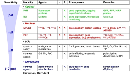
Although most clinical diagnostic imaging studies employ anatomic techniques such as computed tomography (CT) and magnetic resonance (MR) imaging, much of radiology research currently focuses on adapting these conventional methods to physiologic imaging as well as on introducing new techniques and probes for studying processes at the cellular and molecular levels in vivo, i.e. molecular imaging. Molecular imaging promises to provide new methods for the early detection of cancer and support for personalized cancer therapy. Although molecular imaging has been practiced in various incarnations for over 20 years in the context of nuclear medicine, other imaging modalities have only recently been applied to the noninvasive assessment of physiology and molecular events. Nevertheless, there has been sufficient experience with specifically targeted contrast agents and high-resolution techniques for MR imaging and other modalities that we must begin moving these new technologies from the laboratory to the clinic. This brief review outlines several of the more promising areas of pursuit in molecular imaging for oncology with an emphasis on those that show the most immediate likelihood for clinical translation.
International Cancer Imaging Society.
Figures
References
-
- Pomper MG. Molecular imaging: an overview. Acad Radiol. 2001;8:1141–53. - PubMed
-
- Chatziioannou AF. Molecular imaging of small animals with dedicated PET tomographs. Eur J Nucl Med Mol Imaging. 2002;29:98–114. - PubMed
-
- Pomper MG. Can small animal imaging accelerate drug development? J Cell Biochem Suppl. 2002;39:211–20. - PubMed
-
- Doubrovin M, Serganova I, Mayer-Kuckuk P, Ponomarev V, Blasberg RG. Multimodality in vivo molecular-genetic imaging. Bioconjug Chem. 2004;15:1376–88. - PubMed
-
- Guccione S, Yang YS, Shi G, Lee DY, Li KC, Bednarski MD. Functional genomics guided with MR imaging: mouse tumor model study. Radiology. 2003;228:560–8. - PubMed
Publication types
MeSH terms
Substances
Grants and funding
LinkOut - more resources
Full Text Sources
Medical