

The radiology of gastrointestinal stromal tumours (GIST)
- PMID: 16361144
- PMCID: PMC1665232
- DOI: 10.1102/1470-7330.2005.0109
The radiology of gastrointestinal stromal tumours (GIST)
Abstract
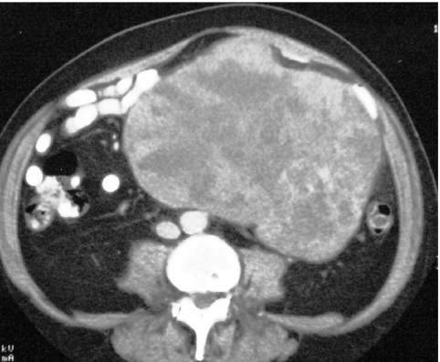
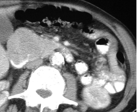
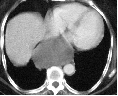
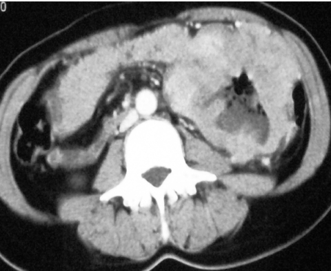
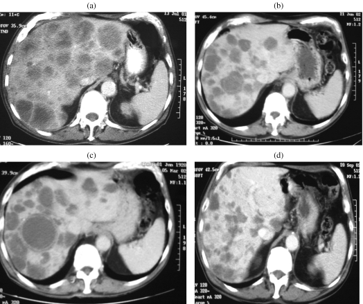
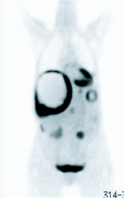
Gastrointestinal stromal tumours (GISTs) comprise a group of smooth muscle mesenchymal alimentary tract tumours of variable malignancy. Recently, the pathophysiology and radiology of these tumours has generated enormous interest following the discovery of a specific, highly effective, chemotherapeutic agent in the form of ST-571 (Imatinib; Glivec, Novartis, Frimley UK). At the time of this review, 106 patients with malignant gastrointestinal stromal tumours seen at the Royal Marsden Hospital have been entered into trials examining the efficacy of varying doses of Imatinib. Burkill et al., also from the Royal Marsden Hospital, have previously reported the distribution, imaging features and pattern of metastatic spread of these tumours (Burkill GJ, Badran M, Al-Muderis O et al. Malignant gastrointestinal stromal tumor: distribution, imaging features, and pattern of metastatic spread. Radiology 2003; 226: 527-32). This new review re-examines the radiological features of GISTs at presentation and well as their changed imaging features following treatment with Imatinib.
International Cancer Imaging Society.
Figures

References
-
- Burkill GJ, Badran M, Al-Muderis O, et al. Malignant gastrointestinal stromal tumor: distribution, imaging features, and pattern of metastatic spread. Radiology. 2003;226:527–32. - PubMed
-
- Sarlamo-Rikala M, Kovatich AJ, Barusevicius A, Miettinen M. CD117: a sensitive marker for gastrointestinal stromal tumors that is more specific than CD34. Mod Pathol. 1998;11:728–34. - PubMed
-
- Fletcher CD, Berman JJ, Corless C, et al. Diagnosis of gastrointestinal stromal tumors: a consensus approach. Hum Pathol. 2002;33:459–65. (review). - PubMed
-
- Miettinen M, Monihan JM, Sarlomo-Rikala M, et al. Gastrointestinal stromal tumors/smooth muscle tumors (GISTs) primary in the omentum and mesentery: clinicopathologic and immunohistochemical study of 26 cases. Am J Surg Pathol. 1999;23:1109–18. - PubMed
Publication types
MeSH terms
Substances
LinkOut - more resources
Full Text Sources
Medical