Review
. 2005 Dec 9;5(1):167-77.
doi: 10.1102/1470-7330.2005.0111.

Imaging of lumps and bumps in the nose: a review of sinonasal tumours

Affiliations
Review

Imaging of lumps and bumps in the nose: a review of sinonasal tumours

Sudip Das et al. Cancer Imaging. .

Abstract

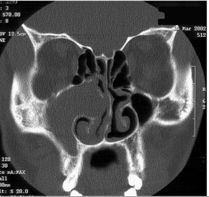
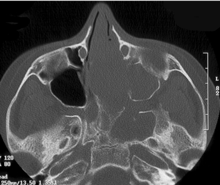
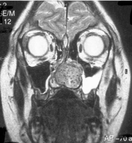
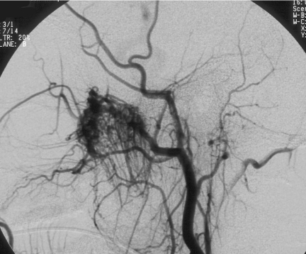
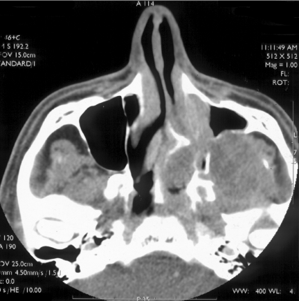
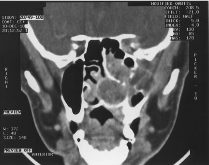
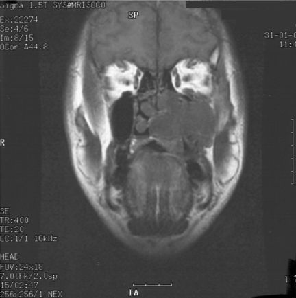
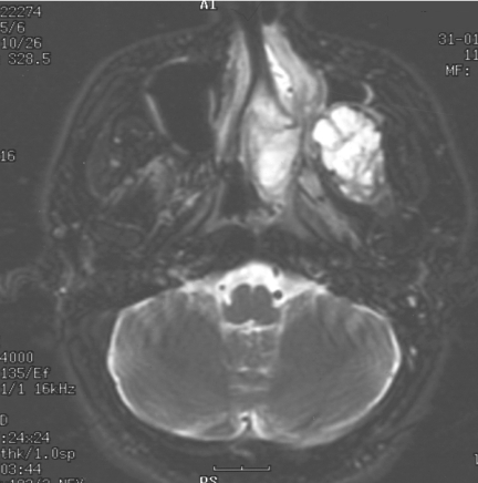
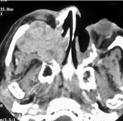
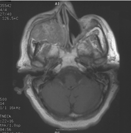
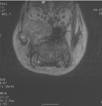
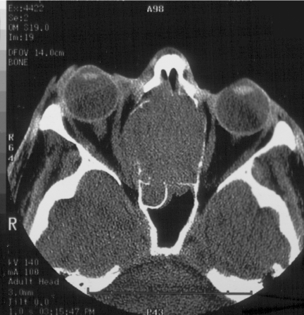
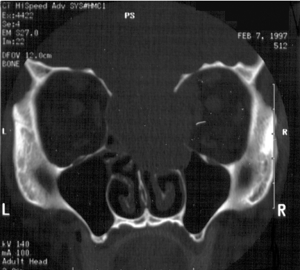
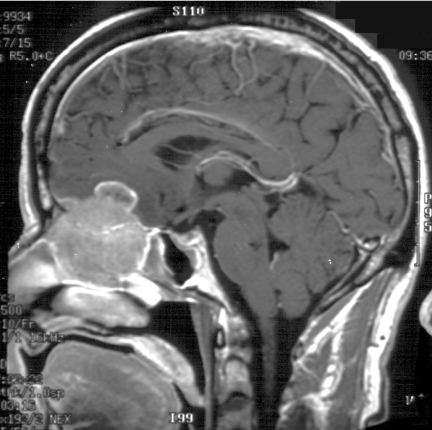
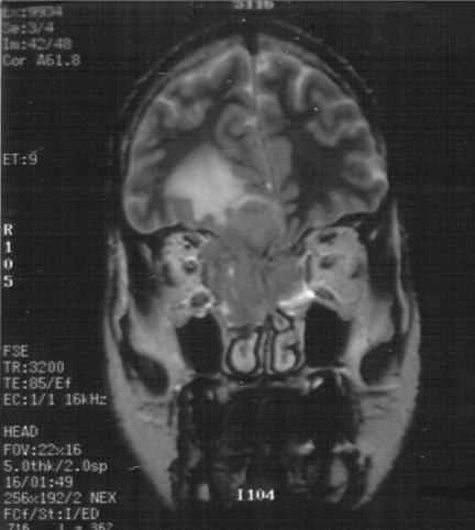
Sinonasal disease is one of the most common clinical head and neck pathologies. The majority of sinonasal pathology is inflammatory with neoplasms comprising approximately 3% of all head and neck tumours. Although sinus tumours are rare, they portend a poor prognosis, often due to advanced disease at diagnosis. Like most neoplasms, early detection improves prognosis, therefore clinicians and radiologists should be aware of features separating tumours from inflammatory sinus disease. This article reviews the anatomy, clinical features, imaging findings, treatment and histopathology of selected sinonasal tumours. Benign neoplasms reviewed include osteoma, inverting papilloma, and juvenile nasal angiofibroma. Malignant neoplasms reviewed include squamous cell carcinoma, the minor salivary gland tumour, adenoid cystic carcinoma, adenocarcinoma, melanoma, lymphoma, and olfactory neuroblastoma (esthesioneuroblastoma).


Figures

Figure 1
Figure 1
Coronal CT scan, bone windows, soft tissue mass obstructing the right osteomeatal unit and complete opacification of the right maxillary antrum. Bony erosion noted along the right maxillary medial wall.
Figure 2
Figure 2
Axial CT bone windows of a juvenile nasal angiofibroma (JNA) completely opacifying the left nasal cavity, enlarging the left pterygopalatine fossa with extension to the left foramen rotundum and vidian canals.
Figure 3
Figure 3
Coronal T2W, MRI of same patient as in Fig. 2, JNA demonstrating multiple small flow voids in the vascular tumour and the utility of MRI in distinguishing tumour from retained secretions in the left maxillary antrum.
Figure 4
Figure 4
Same patient as in Figs 2 and 3. Cerebral angiogram demonstrating marked vascularity of the JNA with feeding vessels from the left internal maxillary artery and branches of the left ascending pharyngeal artery.
Figure 5
Figure 5
Axial CT post-contrast soft tissue windows in a patient with adenoid cystic carcinoma (ACC) extending throughout the left nasal cavity, left maxillary sinus and left pterygoid fossa with enlargement and loss of fat in the left pterygopalatine fossa and left infratemporal fossa. Perineural tumour extension is expected along the V2 branches in pterygopalatine fossa, infraorbital nerve and foramen rotundum.
Figure 6
Figure 6
Coronal CT post-contrast same patient as Fig. 5 with ACC. Demonstrating tumour throughout the left nasal cavity with bony destruction of the left maxillary sinus, loss of fat in the left infratemporal fossa and extension in the left orbital apex along the V2 division.
Figure 7
Figure 7
Same patient as in Figs 5 and 6. Coronal T1 MRI without contrast of ACC filling the left nasal cavity and left maxillary sinus with extension and loss of fat in the left infratemporal fossa. Perineural tumour spread again noted along V2 in the infraorbital foramen.
Figure 8
Figure 8
Same patient as Figs 5–7. Axial T2W MRI in patient with ACC demonstrating increased signal. Adenoid cystic tumours violate the rule of decreased signal on T2W MRI seen with most other cellular tumours, because of their varied histology.
Figure 9
Figure 9
Axial CT post contrast soft tissue windows in patient with adenocarcinoma extending throughout the right nasal cavity, maxillary sinus, and right infratemporal fossa with right intraorbital invasion causing proptosis.
Figure 10
Figure 10
Same patient as in Fig. 9. MRI T1W post-gadolinium of adenocarcinoma showing heterogeneous enhancement of tumour throughout the right sinonasal cavity and maxillary sinus.
Figure 11
Figure 11
Same patient as Figs 9 and 10. Coronal T1W post-gadolinium of adenocarcinoma throughout the right nasal cavity and maxillary sinus with extension into the right infratemporal fossa.
Figure 12
Figure 12
Axial CT bone windows in patient with olfactory neuroblastoma with tumour extension through the right lamina papyrecea causing lateral bowing of the right medial rectus muscle.
Figure 13
Figure 13
Same patient as in Fig. 12; note the right and left sides are reversed on this coronal image. The olfactory neuroblastoma again extends intraorbitally through the lamina papyrecea as well as through the cribiform plate.
Figure 14
Figure 14
Saggital T1W post-gadolinium contrast MRI; same patient as Figs 12 and 13. The olfactory neuroblastoma extends cephalad through the cribiform plate into the anterior cranial fossa and is shown with a cystic component superiorly.
Figure 15
Figure 15
Same patient as Figs 13 and 14. Coronal T2W MRI demonstrating the slightly hypointense olfactory neuroblastoma extending both intraorbitally and intracranially with extensive edema in the right frontal lobe.

Similar articles

Cited by

References

    1. Som P. Tumors and tumorlike conditions of the sinonasal cavity. In: Som P, Bergeron RT, editors. Head and Neck Imaging. 2nd ed. St. Louis: Mosby-Year Book; 1990. pp. 169–227.
    1. Boring CC, Squires TS, Tong T. Cancer statistics. CA Cancer J Clin. 1992;42:19–38. - PubMed
    1. Som PM, Dillon WP, Sze G, et al. Benign and malignant sinonasal lesions with intracranial extension:differentiation with MR imaging. Radiology. 1989;172:763–6. - PubMed
    1. Namdar I, Edelstein DR, Huo J, Lazar A, Kimmelman CP, Soletic R. Management of osteomas of the paranasal sinuses. Am J Rhinol. 1998;12:393–8. - PubMed
    1. Rappaport JM, Attia EL. Pneumocephalus in frontal sinus osteoma: a case report. J Otolaryngol. 1994;23:430–6. - PubMed

MeSH terms

LinkOut - more resources