. 2006 Mar;172(3):1427-39.
doi: 10.1534/genetics.105.051698. Epub 2005 Dec 15.

Glc7-Reg1 phosphatase signals to Yck1,2 casein kinase 1 to regulate transport activity and glucose-induced inactivation of Saccharomyces maltose permease

Affiliations

Glc7-Reg1 phosphatase signals to Yck1,2 casein kinase 1 to regulate transport activity and glucose-induced inactivation of Saccharomyces maltose permease

Nidhi Gadura et al. Genetics. 2006 Mar.

Abstract

The Saccharomyces casein kinase 1 isoforms encoded by the essential gene pair YCK1 and YCK2 control cell growth and morphogenesis and are linked to the endocytosis of several membrane proteins. Here we define roles for the Yck1,2 kinases in Mal61p maltose permease activation and trafficking, using a yck1delta yck2-2(ts) (yck(ts)) strain with conditional Yck activity. Moreover, we provide evidence that Glc7-Reg1 phosphatase acts as an upstream activator of Yck1,2 kinases in a novel signaling pathway that modulates kinase activity in response to carbon source availability. The yck(ts) strain exhibits significantly reduced maltose transport activity despite apparently normal levels and cell surface localization of maltose permease protein. Glucose-induced internalization and rapid loss of maltose transport activity of Mal61/HAp-GFP are not observed in the yck(ts) strain and maltose permease proteolysis is blocked. We show that a reg1delta mutant exhibits a phenotype remarkably similar to that conferred by yck(ts). The reg1delta phenotype is not enhanced in the yck(ts) reg1delta double mutant and is suppressed by increased Yck1,2p dosage. Further, although Yck2p localization and abundance do not change in the reg1delta mutant, Yck1,2 kinase activity, as assayed by glucose-induced HXT1 expression and Mth1 repressor stability, is substantially reduced in the reg1delta strain.


Figures
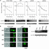
Figure 1.
Figure 1.
Glucose-induced inactivation of maltose permease in yck1Δ yck2ts and YCK1,2 overexpressing strains. (A) Strains LRB906 (YCK1 YCK2) and LRB756 (yck1Δ yck2ts), referred to as yckts, were transformed with plasmids pMAL61/HA, pMAL63, and YEp352, and strain LRB906 was transformed with plasmid YEp352-YCK1 or YEp352-YCK2. Transformants were grown at 24° in selective media plus 2% maltose, harvested by filtration, transferred to YNSG with CHX (30 μg/ml), and standard inactivation protocol followed at 24°. At the indicated times the growth dilution (▵), maltose transport (▪), and relative Mal61/HA protein levels (○) were determined as described in materials and methods. A representative Western blot probed with anti-HA antibody is shown. Western blotting of PGK is shown as the loading control. (B) Strains LRB906 (YCK1 YCK2) and LRB756 (yckts) were transformed with plasmids pMAL61/HA-GFP and pMAL63. Transformants were grown at 24° in synthetic medium with 2% maltose lacking histidine and uracil. Cells were harvested by filtration and transferred to YNSG containing CHX (30 μg/ml) and incubation at 24° with shaking was continued. Culture samples were taken at time zero and every hour over a 3-hr period, gently centrifuged, and GFP localization was visualized by confocal microscropy as described in materials and methods. The images represent a typical result from three independent transformants. (Left) Phase-contrast images. (Right) Fluorescent images. (C) Mal61/HAp expression levels and phosphorylation was determined by Western blotting using total cell extracts prepared from the strains described above in A grown to midlog phase in selective medium plus 2% maltose. Cell extracts were size separated on a 7.5% SDS–PAGE gel and probed with anti-HA and anti-PGK antibody. The arrows indicate the position of the phosphorylated (solid line) and hypophosphorylated (dotted line) species of Mal61/HAp.
Figure 2.
Figure 2.
Comparison of maltose transport activity of yckts and reg1Δ mutant strains. Strains LRB906 (YCK1 YCK2 REG1), LRB756 (yckts), and CMY7000 (reg1Δ) were transformed with plasmids pMAL61/HA and pMAL63 and plasmids YEp352, YEp352-YCK1, YEp352-YCK2, or plasmid DF041 (multi-copy REG1). Transformants were grown at 30° to midlog phase in selective minimal medium plus 2% maltose and maltose transport activity was assayed as described in materials and methods. The error bars indicate the standard deviation from three independent transformants assayed in duplicate.
Figure 3.
Figure 3.
Multicopy YCK2 does not suppress the block in glucose-induced inactivation of maltose permease observed in a doa4Δ mutant. Strain CMY1025 (MAL1 doa4Δ∷HIS3) was transformed with pUN30-MAL61/HA-GFP and the empty vector YEp352 or YEp352-YCK2. Transformants were grown at 30° to midlog phase in synthetic minimal medium lacking tryptophan and uracil plus 2% maltose as carbon source. Cells were harvested by filtration, transferred to YNSG with CHX, and standard inactivation protocol was carried out as described in the legend of Figure 1A. Alternately, cells were observed using confocal fluorescence and phase microscopy at the indicated times following the transfer to YNSG with CHX as described in the legend of Figure 1B. The photos to the left are fluorescent images and those to the right are phase-contrast images.
Figure 4.
Figure 4.
Effects of reg1 deletion and REG1 overexpression on glucose-induced inactivation of maltose permease. (A) Strain CMY7000 (reg1Δ) was transformed with plasmids pUN70-MAL61/HA-GFP and pUN90-MAL63. Transformants were grown at 30° to midlog phase on synthetic medium lacking uracil and histidine plus 2% maltose, harvested by filtration, and transferred to YNSG media with CHX as described in the legend of Figure 1B. Subcellular localization of Mal61/HA-GFP was determined using confocal fluorescence microscopy at the indicated times following transfer to YNSG. (Left) Phase-contrast images. (Right) Fluorescent images. (B) Strain LRB906 (REG1) was transformed with plasmids pRS315-MAL61/HA, pUN90-MAL63, and either pUN70 (vector alone) or plasmid DF041 (multi-copy REG1). Strain CMY7000 (reg1Δ) was transformed with pRS315-MAL61/HA and pUN90-MAL63. Transformants were grown to midlog phase on synthetic medium lacking leucine, histidine, and uracil (for LRB906 transformants) with 2% maltose as the carbon source and glucose-induced inactivation assayed as described in the legend of Figure 1A. (C) The transformed strains described in B were grown in selective medium lacking leucine, histidine, and uracil (for LRB906 transformants) with 2% maltose as the carbon source, harvested, and total cell extracts were prepared and analyzed by Western blotting as described in the legend of Figure 1C. The arrows point to the phosphorylated (solid line) and hypophosphorylated (dotted line) species of Mal61/HA protein.
Figure 5.
Figure 5.
Glucose-induced inactivation of maltose permease in glc7 mutants encoding the catalytic subunit of protein phosphatase type 1. Strains KT1112 (GLC7), KT1636 (glc7-133), KT1639 (glc7-132), KT1967 (glc7-127), KT1638 (glc7-109), and TW267 (glc7-256) were transformed with pRS315-MAL61/HA and pUN90-MAL63. Transformants were grown at 30° to midlog phase on synthetic medium lacking leucine and histidine with 2% maltose. The standard inactivation protocol was followed as described in the legend of Figure 1A.
Figure 6.
Figure 6.
Epistasis analysis places GLC7-REG1 upstream of YCK1 in glucose-induced inactivation of maltose permease. Strains CMY7000 (reg1Δ) and LRB756 (yckts) were transformed with pRS315-MAL61/HA, pUN90-MAL63, and YEp352, pYCK1, or DF041 (REG1). LRB1082 (reg1Δ yckts) was transformed with pRS315-MAL61/HA, pUN90-MAL63, and YEp352 vector. Transformants were grown to midlog phase at 30° on synthetic medium lacking histidine, uracil, and leucine with 2% maltose. The cells were harvested, transferred to YNSG medium with CHX, and the standard inactivation protocol was followed as described in the legend of Figure 1A.(B) Strains LRB906 (YCK1 YCK2 REG1) (WT), LRB756 (yckts), CMY7000 (reg1Δ), and LRB1082 (reg1Δ yckts) were transformed with plasmids pMAL61/HA, pMAL63, and plasmid YEp352 (vector only), pYCK1, or DF041 (REG1), as indicated. Transformants were grown to midlog phase at 30° on synthetic medium lacking histidine, uracil, and leucine with 2% maltose, harvested by filtration, and maltose transport activity was determined as described in materials and methods. The error bars indicate the standard deviation from three independent transformants assayed in duplicate. (C) Phase-contrast images of the strains listed above for B grown to midlog at room temperature; images were taken with Meridian/Olympus IMT-2 confocal microscope using a ×40 lens.
Figure 7.
Figure 7.
reg1Δ blocks glucose-induced HXT1 expression and Mth1 repressor degradation. Strains LRB906 (YCK1 YCK2 REG1), LRB756 (yckts), and CMY7000 (reg1Δ) were transformed with the HXT1-lacZ reporter plasmid pBM3212 (A) or plasmid pBM4560 carrying a 9xMyc-tagged allele of MTH1 expressed from its native promoter (B) (Ozcan et al. 1996). Transformants were grown to midlog at 25° in selective minimal medium lacking leucine (A) or tryptophan (B) plus 2% galactose, harvested by filtration, resuspended in fresh medium plus 2% galactose at 25°, 4% glucose at 25°, or 4% glucose at 30°. (A) Cells were cultured in the indicated medium and temperature for 5 hr, harvested, total cell extracts were prepared, and β-galactosidase levels assayed as described in materials and methods. (B) Cells were cultured in the indicated medium and temperature for 45 min, harvested by filtration, and total cell extracts were prepared for Western blot analysis as described in the legend of Figure 1B except that anti-Myc antibody (Roche Diagnostic) was used to detect the 9xMyc-tagged Mth1p. Equal loading of total-cell extract was confirmed using anti-PGK antibody.

Similar articles

Cited by

References

    1. Babst, M., D. J. Katzmann, E. J. Estepa-Sabal, T. Meerl and S. D. Emr, 2002. Escrt-III: an endosome-associated heterooligomeric protein complex required for mvb sorting. Dev. Cell 3: 271–282. - PubMed
    1. Babu, P., J. D. Bryan, H. R. Panek, S. L. Jordan, B. M. Forbrich et al., 2002. Plasma membrane localization of the Yck2p yeast casein kinase 1 isoform requires the C-terminal extension and secretory pathway function. J. Cell Sci. 115: 4957–4968. - PubMed
    1. Babu, P., R. J. Deschenes and L. C. Robinson, 2004. Akr1p-dependent palmitoylation of Yck2p yeast casein kinase 1 is necessary and sufficient for plasma membrane targeting. J. Biol. Chem. 279: 27138–27147. - PubMed
    1. Baker, S. H., D. L. Frederick, A. Bloecher and K. Tatchell, 1997. Alanine-scanning mutagenesis of protein phosphatase type 1 in the yeast Saccharomyces cerevisiae. Genetics 145: 615–626. - PMC - PubMed
    1. Bonifacino, J. S., and L. M. Traub, 2003. Signals for sorting of transmembrane proteins to endosomes and lysosomes. Annu. Rev. Biochem. 72: 395–447. - PubMed

Publication types

MeSH terms