. 2006 Mar 15;90(6):2062-74.
doi: 10.1529/biophysj.105.071415. Epub 2005 Dec 16.

SNARE-mediated lipid mixing depends on the physical state of the vesicles

Affiliations

SNARE-mediated lipid mixing depends on the physical state of the vesicles

Xiaocheng Chen et al. Biophys J. .

Abstract

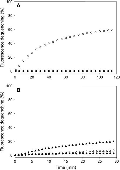
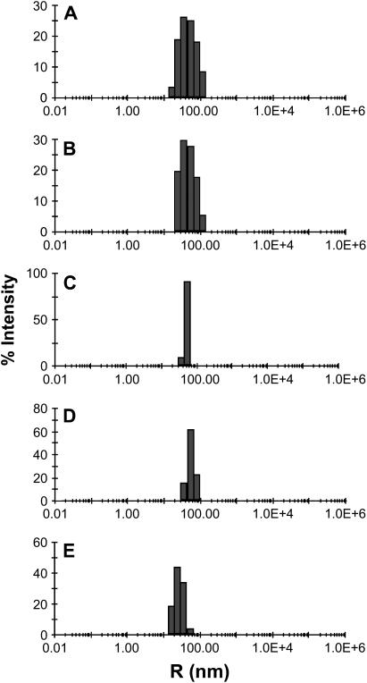
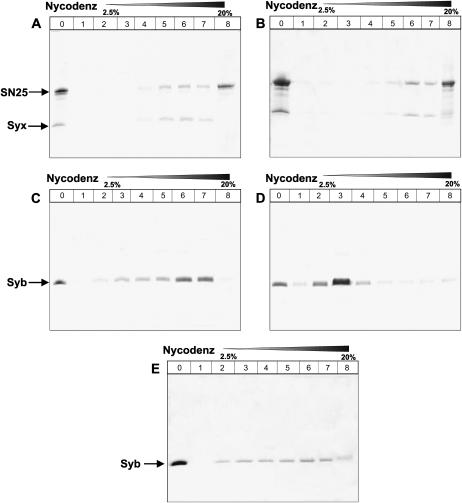
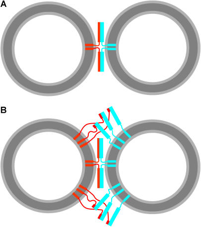
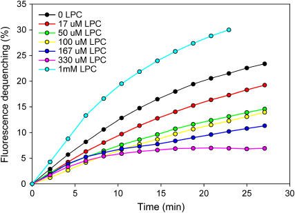
Reconstitution experiments have suggested that N-ethylmaleimide sensitive factor attachment protein receptor (SNARE) proteins constitute a minimal membrane fusion machinery but have yielded contradictory results, and it is unclear whether the mechanism of membrane merger is related to the stalk mechanism that underlies physiological membrane fusion. Here we show that reconstitution of solubilized neuronal SNAREs into preformed 100 nm liposomes (direct method) yields proteoliposomes with more homogeneous sizes and protein densities than the standard reconstitution method involving detergent cosolubilization of proteins and lipids. Standard reconstitutions yield slow but efficient lipid mixing at high protein densities and variable amounts of lipid mixing at moderate protein densities. However, the larger, more homogenous proteoliposomes prepared by the direct method yield almost no lipid mixing at moderate protein densities. These results suggest that the lipid mixing observed for standard reconstitutions is dominated by the physical state of the membrane, perhaps due to populations of small vesicles (or micelles) with high protein densities and curvature stress created upon reconstitution. Accordingly, changing membrane spontaneous curvature by adding lysophospholipids inhibits the lipid mixing observed for standard reconstitutions. Our data indicate that the lipid mixing caused by high SNARE densities and/or curvature stress occurs by a stalk mechanism resembling the mechanism of fusion between biological membranes, but the neuronal SNAREs are largely unable to induce lipid mixing at physiological protein densities and limited curvature stress.


Figures

FIGURE 1
FIGURE 1
Comparison of lipid mixing between synaptobrevin “donor” proteoliposomes and syntaxin/SNAP-25 “acceptor” proteoliposomes prepared by different methods. (A) Lipid mixing measured by fluorescence dequenching using donor proteoliposomes containing 1.5% NBD-DPPE and 1.5% rhodamine-DPPE and a 20:1 lipid/synaptobrevin ratio, and acceptor proteoliposomes with a 150:1 lipid/syntaxin/SNAP-25 ratio (open circles). Both proteoliposome populations were prepared by the standard reconstitution method. NBD fluorescence was normalized using the starting point as 0 and the fluorescence observed after addition of 1% OG as 100. The solid circles represent an analogous experiment performed with protein-free liposomes as acceptor vesicles. (B) Analogous experiments performed with synaptobrevin and syntaxin/SNAP-25 proteoliposomes prepared by the standard method with 160:1 and 150:1 lipid/protein ratios, respectively (solid and open triangles, which represent experiments performed under the same conditions but with different preparations), or with synaptobrevin and syntaxin/SNAP-25 proteoliposomes prepared by the direct method with 185:1 and 200:1 lipid/protein ratios, respectively (solid circles). All experiments were performed with a 1 mM total lipid concentration.
FIGURE 2
FIGURE 2
The direct method yields proteoliposomes with more homogeneous size distributions. The diagrams show DLS analyses of the proteoliposomes used for the lipid mixing assays shown in Fig. 1 but diluted 100-fold (10 μM final lipid concentration). (A and E) Synaptobrevin proteoliposomes prepared by the standard method with a 160:1 lipid/protein ratio (A) or a 20:1 lipid/protein ratio (E). (B) Syntaxin/SNAP-25 proteoliposomes prepared by the standard method with a 150:1 lipid/protein ratio. (C and D) Synaptobrevin (C) and syntaxin/SNAP-25 (D) proteoliposomes prepared by the direct method with lipid/protein ratios of 185:1 and 200:1, respectively.
FIGURE 3
FIGURE 3
Direct method yields synaptobrevin proteoliposomes with more homogeneous protein densities. (AE) Nycodenz gradient flotation analyses of proteoliposomes containing neuronal SNAREs are shown. (A,C, and E) Proteoliposomes prepared by the standard method and containing syntaxin/SNAP-25 (A) or synaptobrevin (C and E) at 150:1 and 160:1 initial lipid/protein ratios, respectively. Panels C and E represent two experiments performed with different preparations under the same conditions. (B and D) Proteoliposomes prepared by the direct method and containing syntaxin/SNAP-25 (B) or synaptobrevin (D) at 185:1 and 200:1 initial lipid/protein ratios, respectively. Proteoliposomes were floated on a gradient containing Nycodenz concentrations that ranged from 2.5% to 40%. Fractions were pooled and analyzed by SDS-PAGE followed by Coomassie blue staining. Lane 0 corresponds to the starting material, and lanes 1–7 correspond to fractions containing 2.5–20% Nycodenz, where all proteoliposomes were found. Lane 8 corresponds to the bottom of the gradient (30–40% Nycodenz), which only contained unincorporated proteins. The volume of this layer used for SDS-PAGE analysis was 10-fold larger than those used for fractions 1–7 to facilitate detection of small amounts of unincorporated protein (see Materials and Methods).
FIGURE 4
FIGURE 4
How many SNARE complexes induce lipid mixing? (A) Model of two fully assembled SNARE complexes located between the most proximal regions of two vesicles. The model was drawn approximately to scale to represent the relative sizes of the SNARE complexes and 40 nm vesicles and to illustrate that SNARE complex assembly should bring the opposing membranes within 2–3 nm, but it is unclear whether this is sufficient for membrane merger. (B) Model analogous to A but including additional SNARE complexes that are initiating assembly at their N-termini with the rest of their SNARE motifs unstructured. The model is intended to illustrate the possibility that massive formation of SNARE complexes between more distal parts of the two membranes might actually induce lipid mixing rather than formation of a few SNARE complexes in the proximal intermembrane space. In panels A and B, synaptobrevin is colored in red and syntaxin/SNAP-25 heterodimers in cyan. Curves with arbitrary shapes indicate unstructured regions (i.e., the SNARE motif of free synaptobrevin and the linker sequences between the TM regions and the SNARE motifs of syntaxin and synaptobrevin). Thin rectangles represent individual helices (i.e., TM regions and the synaptobrevin SNARE motif upon partial or full assembly of SNARE complexes), and wide rectangles represent the helix bundle formed by the syntaxin and SNAP-25 SNARE motifs.
FIGURE 5
FIGURE 5
No significant docking is observed during lipid mixing reactions mediated by the neuronal SNAREs. The figure shows a cryo-EM image of a mixture of donor and acceptor vesicles prepared under the same conditions to those used for the lipid mixing assays shown in Fig. 1 A. The sample was fast-frozen a few minutes after mixing donor and acceptor proteoliposomes. The scale bar corresponds to 100 nm.
FIGURE 6
FIGURE 6
Analysis of fluorescein leakage of neuronal SNARE proteoliposomes. (A and B) Fluorescence spectra at 37°C as a function of time of samples of synaptobrevin (A) and syntaxin/SNAP-25 (B) proteoliposomes obtained by the standard method with analogous protein densities to those used for the lipid mixing assays of Fig. 1 A and with trapped fluorescein (100 mM). The top curve represents the fluorescence spectrum obtained upon addition of 1% OG after 2 h of incubation. (C) Time dependence of the fluorescence intensity at 518 nm observed in the experiments of panels A and B, expressed as percentage of the fluorescence intensity obtained after liposome lysis with 1% OG (circles, synaptobrevin liposomes; triangles, syntaxin/SNAP-25 liposomes). (D) Fluorescence spectra at 37°C as a function of time of a mixture of synaptobrevin vesicles and syntaxin/SNAP-25 vesicles analogous to those used in panels A and B. (E) Time dependence of the fluorescence intensity at 518 nm for the experiment shown in panel D. (F) SDS-PAGE analysis of the mixture used for the experiments in (D and E) at 0, 1, and 2 h (left lanes) and of a comparable mixture with proteoliposomes prepared without trapped fluorescein. Note the appearance of SDS-resistant SNARE complexes in the latter but not in the former experiment.
FIGURE 7
FIGURE 7
Lysophosphatidylcholine inhibits SNARE-mediated lipid mixing. Lipid mixing assays were performed and monitored as in Fig. 1 A but with different additions of oleoyl LPC at the start of the reaction. The final concentrations of oleoyl LPC are color coded and indicated in the inset.

References

    1. Pelham, H. R. 1999. SNAREs and the secretory pathway—lessons from yeast. Exp. Cell Res. 247:1–8. - PubMed
    1. Chen, Y. A., and R. H. Scheller. 2001. SNARE-mediated membrane fusion. Nat. Rev. Mol. Cell Biol. 2:98–106. - PubMed
    1. Rizo, J., and T. C. Sudhof. 2002. Snares and munc18 in synaptic vesicle fusion. Nat. Rev. Neurosci. 3:641–653. - PubMed
    1. Jahn, R., T. Lang, and T. C. Sudhof. 2003. Membrane fusion. Cell. 112:519–533. - PubMed
    1. Schiavo, G., M. Matteoli, and C. Montecucco. 2000. Neurotoxins affecting neuroexocytosis. Physiol. Rev. 80:717–766. - PubMed

Publication types