. 2005 Dec 27;102(52):18932-7.
doi: 10.1073/pnas.0509469102. Epub 2005 Dec 16.

Structural basis for conductance by the archaeal aquaporin AqpM at 1.68 A

Affiliations

Structural basis for conductance by the archaeal aquaporin AqpM at 1.68 A

John K Lee et al. Proc Natl Acad Sci U S A. .

Abstract

To explore the structural basis of the unique selectivity spectrum and conductance of the transmembrane channel protein AqpM from the archaeon Methanothermobacter marburgensis, we determined the structure of AqpM to 1.68-A resolution by x-ray crystallography. The structure establishes AqpM as being in a unique subdivision between the two major subdivisions of aquaporins, the water-selective aquaporins, and the water-plus-glycerol-conducting aquaglyceroporins. In AqpM, isoleucine replaces a key histidine residue found in the lumen of water channels, which becomes a glycine residue in aquaglyceroporins. As a result of this and other side-chain substituents in the walls of the channel, the channel is intermediate in size and exhibits differentially tuned electrostatics when compared with the other subfamilies.


Figures

Fig. 1.
Fig. 1.
Monomer and tetramer view of AqpM. (A) AqpM monomer viewed parallel to the plane of the membrane. The six transmembrane (M1–M2, M4–M6, and M8) and two half-membrane (M3 and M7) spanning helices are labeled M1–M8. The glycerol and water molecules found in the channel are represented as spheres. The two OG detergent molecules identified in the structure are represented as transparent spheres. The channel is readily identifiable by the line of water and glycerol molecules coordinated inside the channel. (B) The AqpM tetramer viewed down the 4-fold axis of the tetramer from the extracellular aspect of the membrane. Four residues that form the selectivity filter are shown in stick rendering. (C) Stereoview of the sigma-weighted 2 FoFc electron density at the selectivity filter. The map is contoured at 1.2 σ. The figure was made with molscript (25), raster3d (26), and pymol (27).
Fig. 2.
Fig. 2.
AqpM channel architecture. Stereoview of the channel with potential hydrogen bonds to the waters (HOH1–H4) and glycerols (G1–G3) shown in gray dotted lines. Helices of M3 and M7 are rendered in cartoon representation, while the loop portion residues are represented as sticks. The main-chain carbonyl oxygens of the two loops form the conserved ladder-like structure of hydrogen-bond acceptors that line the channel. N82 and N199 are the asparagines that make up the canonical aquaporin NPA motifs.
Fig. 3.
Fig. 3.
Aquaporin selectivity filters. (A) The residues of the selectivity filter of AqpM (R202, F62, S196, and I187) are depicted with the water molecule (HOH2) coordinated at the center of the filter. The potential hydrogen bonds from HOH2 to S196 and R202 are depicted as a blue dotted line. (B) The sequence alignment of six aquaporins. I187 from AqpM and corresponding residues in the other aquaporins are highlighted in the red box. In water-selective aquaporins, represented here by Aqp1 and AqpZ, a conserved histidine residue occupies this position. In aquaglyceroporins such as GlpF, a glycine residue occupies this position. Similar to AqpM, the two other archaeal aquaporins [A. fulgidus aquaporin (AfAqp) and Methanosarcina barkeri aquaporin (MbAqp)] possess aliphatic residues at this position. SaAqp, S. acidocaldarius aquaporin. MmAqp, Methanococcus maripaludis aquaporin. (C and D) The selectivity filters of Aqp1 (C) and GlpF (D) highlighting the similarity and differences between an aquaporin and aquaglyceroporin. A glycerol molecule is seen only in the selectivity filter of GlpF (D).
Fig. 4.
Fig. 4.
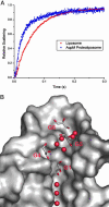
Water conductance by AqpM and water and glycerol in the vestibule of the channel. (A) Water permeability of liposome-reconstituted AqpM. Liposomes reconstituted with purified AqpM or control liposomes were mixed at 12.2°C with an equal volume of hyperosmolar solution (570 mM sucrose). The reduction in vesicular volume caused by water efflux, resulting in an increase in light scattering, was observed in a stopped-flow apparatus for 0.5 s. The data were normalized between zero and unity and fitted to an exponential rise to the maximal value curve. Osmotic water permeabilities constants were calculated as follows: Pf(liposomes) = 15.5 ± 0.7 μm/s, Pf(AqpM proteoliposomes) = 60.5 ± 3 μm/s. (B) Shown is the surface rendering of AqpM with helices 3, 4, 7, and 8 removed to highlight the channel and vestibule. Three glycerol (G4–G6) and five water molecules can be seen in the pocket of the vestibule on the extracellular side of the channel. With its hydrophobic backbone facing the greasy portion of the channel, G4 is approximately in the optimal orientation before entering the narrow portion of the channel.

Similar articles

Cited by

References

    1. Preston, G. M., Carroll, T. P., Guggino, W. B. & Agre, P. (1992) Science 256, 385–387. - PubMed
    1. Fu, D., Libson, A., Miercke, L. J., Weitzman, C., Nollert, P., Krucinski, J. & Stroud, R. M. (2000) Science 290, 481–486. - PubMed
    1. Murata, K., Mitsuoka, K., Hirai, T., Walz, T., Agre, P., Heymann, J. B., Engel, A. & Fujiyoshi, Y. (2000) Nature 407, 599–605. - PubMed
    1. Sui, H., Han, B. G., Lee, J. K., Walian, P. & Jap, B. K. (2001) Nature 414, 872–878. - PubMed
    1. Savage, D. F., Egea, P. F., Robles-Colmenares, Y., O'Connell, J. D., 3rd & Stroud, R. M. (2003) PloS Biol. 1, 334–340. - PMC - PubMed

Publication types

MeSH terms