Role of petal-specific orcinol O-methyltransferases in the evolution of rose scent
- PMID: 16361520
- PMCID: PMC1326028
- DOI: 10.1104/pp.105.070961
Role of petal-specific orcinol O-methyltransferases in the evolution of rose scent
Abstract
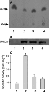
Orcinol O-methyltransferase (OOMT) 1 and 2 catalyze the last two steps of the biosynthetic pathway leading to the phenolic methyl ether 3,5-dimethoxytoluene (DMT), the major scent compound of many rose (Rosa x hybrida) varieties. Modern roses are descended from both European and Chinese species, the latter being producers of phenolic methyl ethers but not the former. Here we investigated why phenolic methyl ether production occurs in some but not all rose varieties. In DMT-producing varieties, OOMTs were shown to be localized specifically in the petal, predominantly in the adaxial epidermal cells. In these cells, OOMTs become increasingly associated with membranes during petal development, suggesting that the scent biosynthesis pathway catalyzed by these enzymes may be directly linked to the cells' secretory machinery. OOMT gene sequences were detected in two non-DMT-producing rose species of European origin, but no mRNA transcripts were detected, and these varieties lacked both OOMT protein and enzyme activity. These data indicate that up-regulation of OOMT gene expression may have been a critical step in the evolution of scent production in roses.
Figures


Similar articles
-
Scent evolution in Chinese roses.Proc Natl Acad Sci U S A. 2008 Apr 15;105(15):5927-32. doi: 10.1073/pnas.0711551105. Epub 2008 Apr 14. Proc Natl Acad Sci U S A. 2008. PMID: 18413608 Free PMC article.
-
Biosynthesis of the major scent components 3,5-dimethoxytoluene and 1,3,5-trimethoxybenzene by novel rose O-methyltransferases.FEBS Lett. 2002 Jul 17;523(1-3):113-8. doi: 10.1016/s0014-5793(02)02956-3. FEBS Lett. 2002. PMID: 12123815
-
O-methyltransferases involved in the biosynthesis of volatile phenolic derivatives in rose petals.Plant Physiol. 2002 Aug;129(4):1899-907. doi: 10.1104/pp.005330. Plant Physiol. 2002. PMID: 12177504 Free PMC article.
-
Floral benzenoid carboxyl methyltransferases: from in vitro to in planta function.Phytochemistry. 2005 Jun;66(11):1211-30. doi: 10.1016/j.phytochem.2005.03.031. Phytochemistry. 2005. PMID: 15946712 Free PMC article. Review.
-
Flower colour and cytochromes P450.Philos Trans R Soc Lond B Biol Sci. 2013 Jan 6;368(1612):20120432. doi: 10.1098/rstb.2012.0432. Print 2013 Feb 19. Philos Trans R Soc Lond B Biol Sci. 2013. PMID: 23297355 Free PMC article. Review.
Cited by
-
Identification of white campion (Silene latifolia) guaiacol O-methyltransferase involved in the biosynthesis of veratrole, a key volatile for pollinator attraction.BMC Plant Biol. 2012 Aug 31;12:158. doi: 10.1186/1471-2229-12-158. BMC Plant Biol. 2012. PMID: 22937972 Free PMC article.
-
Genetic and Biochemical Aspects of Floral Scents in Roses.Int J Mol Sci. 2022 Jul 20;23(14):8014. doi: 10.3390/ijms23148014. Int J Mol Sci. 2022. PMID: 35887360 Free PMC article. Review.
-
Phenotypic Space and Variation of Floral Scent Profiles during Late Flower Development in Antirrhinum.Front Plant Sci. 2016 Dec 21;7:1903. doi: 10.3389/fpls.2016.01903. eCollection 2016. Front Plant Sci. 2016. PMID: 28066463 Free PMC article.
-
Genetics and genomics of flower initiation and development in roses.J Exp Bot. 2013 Feb;64(4):847-57. doi: 10.1093/jxb/ers387. Epub 2013 Jan 29. J Exp Bot. 2013. PMID: 23364936 Free PMC article. Review.
-
Regulators of floral fragrance production and their target genes in petunia are not exclusively active in the epidermal cells of petals.J Exp Bot. 2012 May;63(8):3157-71. doi: 10.1093/jxb/ers034. Epub 2012 Feb 15. J Exp Bot. 2012. PMID: 22345641 Free PMC article.
References
-
- Boevink P, Oparka K, Santa Cruz S, Martin B, Betteridge A, Hawes C (1998) Stacks on tracks: the plant Golgi apparatus traffics on an actin/ER network. Plant J 15: 441–447 - PubMed
-
- Brandizzi F, Irons SL, Johansen J, Kotzer A, Neumann U (2004) GFP is the way to glow: bioimaging of the plant endomembrane system. J Microsc 214: 138–158 - PubMed
Publication types
MeSH terms
Substances
Associated data
- Actions
- Actions
- Actions
- Actions
- Actions
- Actions
- Actions
- Actions
- Actions
- Actions
- Actions
- Actions
- Actions
- Actions
- Actions
LinkOut - more resources
Full Text Sources
Molecular Biology Databases

