. 2006 Jan;140(1):81-90.
doi: 10.1104/pp.105.066464. Epub 2005 Dec 16.

Lectin receptor kinases participate in protein-protein interactions to mediate plasma membrane-cell wall adhesions in Arabidopsis

Affiliations

Lectin receptor kinases participate in protein-protein interactions to mediate plasma membrane-cell wall adhesions in Arabidopsis

Anne Gouget et al. Plant Physiol. 2006 Jan.

Abstract

Interactions between plant cell walls and plasma membranes are essential for cells to function properly, but the molecules that mediate the structural continuity between wall and membrane are unknown. Some of these interactions, which are visualized upon tissue plasmolysis in Arabidopsis (Arabidopsis thaliana), are disrupted by the RGD (arginine-glycine-aspartic acid) tripeptide sequence, a characteristic cell adhesion motif in mammals. In planta induced-O (IPI-O) is an RGD-containing protein from the plant pathogen Phytophthora infestans that can disrupt cell wall-plasma membrane adhesions through its RGD motif. To identify peptide sequences that specifically bind the RGD motif of the IPI-O protein and potentially play a role in receptor recognition, we screened a heptamer peptide library displayed in a filamentous phage and selected two peptides acting as inhibitors of the plasma membrane RGD-binding activity of Arabidopsis. Moreover, the two peptides also disrupted cell wall-plasma membrane adhesions. Sequence comparison of the RGD-binding peptides with the Arabidopsis proteome revealed 12 proteins containing amino acid sequences in their extracellular domains common with the two RGD-binding peptides. Eight belong to the receptor-like kinase family, four of which have a lectin-like extracellular domain. The lectin domain of one of these, At5g60300, recognized the RGD motif both in peptides and proteins. These results imply that lectin receptor kinases are involved in protein-protein interactions with RGD-containing proteins as potential ligands, and play a structural and signaling role at the plant cell surfaces.


Figures
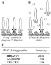
Figure 1.
Figure 1.
Selection of phages displaying random heptamer peptides that interact with the RGD sequence of the IPI-O protein. A, The library of phage-displayed peptides preincubated with BSA was incubated into microtiter plates coated with the IPI-O protein (lobster claw; first step). B, After extensive washing to remove unbound phages, competition with an RGD-containing peptide (broken square) was carried out to release a specific subset of the bound phages (second step). C, After a single round of bio-panning selection, 36 phages were randomly picked and the inserts were sequenced. The three different heptapeptides obtained are shown with their frequency.
Figure 2.
Figure 2.
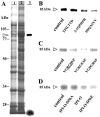
Photoaffinity labeling of plasma membrane proteins from Arabidopsis with an azido RGD heptapeptide derivative, and effect of the RGD-binding heptapeptides selected by phage display. Plasma membrane proteins were separated in SDS-11% polyacrylamide gels after photolysis of the radioiodinated RGD-photoaffinity probe as described in “Materials and Methods.” Autoradiography revealed label associated with a protein of 80 kD. To quantify cross-linking, gel slices corresponding to the 80-kD polypeptide were excised and γ counted. The values of remaining label are indicated below each band as percentages of the control that was set at 100% in each independent experiment. Fifty micrograms of protein were deposited in each lane. A, Competition by isolated RGD-binding heptapeptides (IHQASYY, TPILTTD, AAQPHPR), a c-myc peptide with unrelated sequence (EQKLISEEDL; each peptide was applied at the concentration of 100 μm), and wild-type and mutated (-D56A, -D56E) MBP-IPI-O proteins (each protein was applied at the concentration of 0.3 μm). B, Competition by the two active RGD-binding heptapeptides (AAQPHPR and IHQASYY) and derived tetrapeptides. Each peptide was applied at the concentration of 100 μm.
Figure 3.
Figure 3.
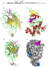
Schematic representations of the lectin-like receptor kinase encoded by At5g60300. A, The NH2-terminal signal peptide is displayed as a gray box: It is presumably absent in the mature protein. The legume lectin domain is composed of 13 β-strands (black boxes) that form antiparallel β-sheets. The transmembrane domain is shown as a gray box (TM). The 12 subdomains of the kinase region are numbered in Roman numerals (I–XI). The amino acid sequences found in the isolated phage-displayed peptides are given above. B, Ribbon diagram of the modeled lectin-like domain of At5g60300 showing the overall β-sandwich fold organized in a flattened back face of a six-stranded β-sheet connected by turns and loops to a curved front face of a seven-stranded β-sheet. β-Strands are represented by yellow arrows, and coil structures are colored green. The additional extended loop (open star) contains a short α-helical stretch (colored red). Residues Ser-152, Tyr-153, and Tyr-154 (magenta ball-and-sticks) involved in the recognition of the RGD motif of IPI-O are located in an exposed loop connecting strand β8 to β9. The location of the putative carbohydrate-binding site is indicated (★). Strand β1 could participate in the dimerization of the lectin-like domain of At5g60300. C, Ribbon diagram of the modeled Ser/Thr-kinase domain of At5g30600 built up from a small β-rich N-terminal lobe (top part) connected to a large α-helical C-terminal lobe (bottom part). ATP (magenta CPK, Corey-Pauling-Koltun) is docked into the catalytic cavity located at the interface of the two lobes. D, Sagital view of the modeled lectin-like domain showing the location of the Asn residues belonging to the possibly glycosylated putative N-glycosylation sites. E, Molecular surface of the modeled lectin-like domain showing the location of the RGD-binding site S-Y-Y (colored magenta), the putative N-glycosylation sites (colored blue), and the apparently inactive carbohydrate-binding cavity (★). The lectin-like domain is similarly oriented in D and E.
Figure 4.
Figure 4.
Specificity of the photoaffinity labeling of crude extracts obtained from an E. coli strain expressing the lectin domain of At5g60300. A, Lanes 1 and 2 are electrophoregrams obtained from total proteins of E. coli containing either an empty pTYB12 vector (lane 1) or a pTYB12 vector with a partial open reading frame of At5g60300 encoding the extracellular lectin domain as insert (lane 2): The recombinant protein appeared as a major band at a molecular mass of 85 kD (arrowhead). Lane 3 is the labeling pattern observed after autoradiography when the radioiodinated RGD-photoaffinity probe was photolysed as described in “Materials and Methods.” Ten micrograms were deposited in lanes 1 and 2, 0.8 μg of protein in lane 3. B, Labeling patterns and competition by RGD-binding peptides: Each peptide was applied at the concentration of 200 μm. A total of 0.8 μg of recombinant protein was deposited in each lane. C, Labeling patterns and competition by peptides containing a full or modified RGD motif: Each peptide was applied at the concentration of 200 μm. A total of 0.8 μg of protein was deposited in each lane. D, Labeling patterns and competition by the wild-type and mutant MBP-IPI-O proteins: Each protein was applied at the concentration of 0.3 μm. A total of 0.8 μg of protein was deposited in each lane.
Figure 5.
Figure 5.
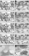
Plasma membrane-cell wall adhesions in Arabidopsis hypocotyls. Hypocotyls from 8-d-old etiolated seedlings were prepared and stained with neutral red as described in “Materials and Methods.” Upon addition of 0.4 m CaCl2, time courses of plasmolysis were observed for 10 min. Plasmolysis was induced in the absence of additives. A and B show a portion of entire hypocotyls. A, Control series without additives: The plasmolysed cells revealed plasma membrane-cell wall adhesions (arrows) and concave forms of plasmolysis (triangles). B, RGD-binding peptides (1 mm IHQASYY and AAQPHPR) were added to the plasmolysed hypocotyls: The plasma membrane quickly separates from the wall to make spherical protoplasts and convex forms of plasmolysis. C and D are enlarged images to show wall-to-membrane adhesions and concave forms of plasmolysis (C), as well as convex forms of plasmolysis in the presence of IHQASYY and AAQPHPR peptides (D).

Similar articles

Cited by

References

    1. Arabidopsis Genome Initiative (2000) Analysis of the genome sequence of the flowering plant Arabidopsis thaliana. Nature 408: 796–815 - PubMed
    1. Bardy N, Carrasco A, Galaud JP, Pont-Lezica R, Canut H (1998) Free-flow electrophoresis for fractionation of Arabidopsis membranes. Electrophoresis 19: 1145–1153 - PubMed
    1. Barre A, Hervé C, Lescure B, Rougé P (2002) Lectin receptor kinases in plants. CRC Crit Rev Plant Sci 21: 379–399
    1. Bateman A, Coin L, Durbin R, Finn RD, Hollich V, Griffiths-Jones S, Khanna A, Marshall M, Moxon S, Sonnhammer EL, et al (2004) The Pfam protein families database. Nucleic Acids Res 32: D138–D141 - PMC - PubMed
    1. Berger F, Taylor A, Brownlee C (1994) Cell fate determination by the cell wall in early Fucus development. Science 263: 1421–1423 - PubMed

Publication types

MeSH terms

Associated data