

Ultrasound biomicroscopy of Chinese eyes with iridocorneal endothelial syndrome
- PMID: 16361670
- PMCID: PMC1856895
- DOI: 10.1136/bjo.2005.074864
Ultrasound biomicroscopy of Chinese eyes with iridocorneal endothelial syndrome
Abstract
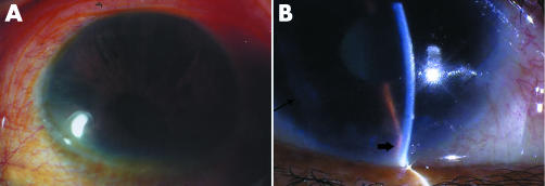
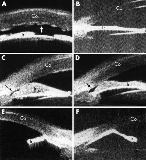
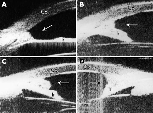
Aim: To document the ultrasound biomicroscopic (UBM) findings in Chinese patients with iridocorneal endothelial (ICE) syndrome.
Methods: 21 patients with ICE syndrome and 15 normal subjects underwent UBM. UBM findings of anterior segment were compared between normal subjects and three clinical types of ICE syndrome: progressive iris atrophy (PIA), Chandler's syndrome (CS), and Cogan-Reese syndrome (CRS).
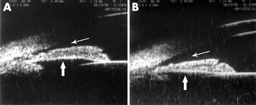
Results: Central anterior chamber depth was significantly less in patients with ICE syndrome (2.25 (SD 0.32) mm) than in normal subjects (2.76 (0.32) mm). Peripheral anterior synechiae were observed in all the ICE patients by UBM. Three out of four CRS subjects showed an "arborised" shape of iridocorneal angle. Two eyes out of 10 with CS presented bridge-shaped synechiae. A membrane-like mound was observed in iridocorneal angle in two patients: one with CRS and one with CS. UBM was found to be more effective in detecting peripheral anterior synechiae (PAS) and iris atrophy than slit lamp microscopy and gonioscopy, mainly because of corneal oedema in patients with CS. Four out of 11 patients with unilateral ICE syndrome had shallow or closed anterior chamber angles in their fellow eyes. Two of them successfully responded to laser peripheral iridotomy.
Conclusions: UBM is an effective method to reveal the anterior segment features and provides a useful tool in the diagnosis of ICE syndrome. Different subtypes of ICE syndrome may have different UBM manifestations. UBM can help to identify angle closure in the fellow eye of unilateral ICE syndromes.
Conflict of interest statement
Competing interest statement: No financial interests related to the manuscript.
References
-
- Shields M B. Progressive essential iris atrophy, Chandler's syndrome, and the iris nevus (Cogan‐Reese) syndrome: a spectrum of disease. Surv Ophthalmol 1979243–20. - PubMed
-
- Kidd M, Hetherington J, Magee S. Surgical results in iridocorneal endothelial syndrome. Arch Ophthalmol 1988106199–201. - PubMed
-
- Kim D K, Aslanides I M, Schmidt C M., Jret al Long‐term outcome of aqueous shunt surgery in ten patients with iridocorneal endothelial syndrome. Ophthalmology 19991061030–1034. - PubMed
Publication types
MeSH terms
LinkOut - more resources
Full Text Sources