Review
. 2005 Dec;20(6):913-25.
doi: 10.3346/jkms.2005.20.6.913.

Diagnosis and treatment of nontuberculous mycobacterial pulmonary diseases: a Korean perspective

Affiliations
Review

Diagnosis and treatment of nontuberculous mycobacterial pulmonary diseases: a Korean perspective

Won-Jung Koh et al. J Korean Med Sci. 2005 Dec.

Abstract

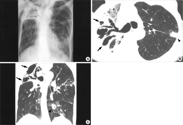
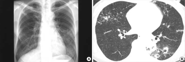
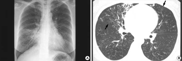
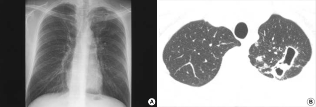
The incidence of pulmonary disease caused by nontuberculous mycobacteria (NTM) appears to be increasing worldwide. In Korea, M. avium complex and M. abscessus account for most of the pathogens encountered, whilst M. kansasii is a relatively uncommon cause of NTM pulmonary diseases. NTM pulmonary disease is highly complex in terms of its clinical presentation and management. Because its clinical features are indistinguishable from those of pulmonary tuberculosis and NTMs are ubiquitous in the environment, the isolation and identification of causative organisms are mandatory for diagnosis, and some specific diagnostic criteria have been proposed. The treatment of NTM pulmonary disease depends on the infecting species, but decisions concerning the institution of treatment are never easy. Treatment requires the use of multiple drugs for 18 to 24 months. Thus, treatment is expensive, often has significant side effects, and is frequently not curative. Therefore, clinicians should be confident that there is sufficient pathology to warrant prolonged, multidrug treatment regimens. In all of the situations, outcomes can be best optimized only when clinicians, radiologists, and laboratories work cooperatively.


Figures

Fig. 1
Fig. 1
M. intracellulare pulmonary disease of the upper lobe cavitary form in a 73-yr-old man. Chest radiograph shows parenchymal opacity which is containing multiple cavities in the right upper lobe. Also note nodular lesions in left middle and lower lung zones (A). Transaxial lung window CT image (2.5-mm section thickness, 70 mA) obtained at the level of the right upper lobar bronchus shows dilated bronchi in an opacified right upper lobe. Also note associated multiple cavitary lesions (arrows). A lobular consolidative lesion (arrowhead) is seen in the left upper lobe (B). Coronal reformation (2.0-mm section thickness) image demonstrates dilated bronchi and multiple thin walled cavities (arrows) in the right upper lobe. Also note multiple variable-sized nodules and consolidation (arrowhead) in the left lung (C).
Fig. 2
Fig. 2
M. intracellulare pulmonary disease of the nodular bronchiectatic form in a 63-yr-old woman. Chest radiograph shows a multifocal patchy distribution of small nodular clusters in both lungs (A). Transaxial lung window CT image (2.5-mm section thickness, 70 mA) obtained at the level of basal trunk (B) show small centrilobular nodules and bronchiectasis in the right middle lobe and in the lingular division of the left upper lobe. Also note small cavitating nodules and lobular consolidation in the left lower lobe.
Fig. 3
Fig. 3
M. abscessus pulmonary disease in a 49-yr-old woman. Chest radiograph shows multifocal patchy areas of small nodular clusters in both lungs. Also note parenchymal opacity in the right middle lobe (A). Transaxial lung window CT image (2.5-mm section thickness, 70 mA) obtained at the basal trunk (B) show bronchiectasis and small centrilobular nodules or tree-in-bud opacities (arrows) in the right middle lobe and in the lingular division of the left upper lobe. Also note bronchiolitis of small centrilobular nodules and tree-in-bud opacities in both lower lobes.
Fig. 4
Fig. 4
M. kansasii pulmonary disease in a 30-yr-old man. Chest radiograph shows cavitary and small nodular lesions in the left upper lobe (A). Transaxial lung window CT thin-section images (1.0-mm section thickness, 170 mA) obtained at the thoracic inlet level show multiple thin-walled cavities in the left upper lobe. Also note small nodular lesions and tree-in-bud opacities in the left upper lobe (B).

References

    1. Hale YM, Pfyffer GE, Salfinger M. Laboratory diagnosis of mycobacterial infections: new tools and lessons learned. Clin Infect Dis. 2001;33:834–846. - PubMed
    1. American Thoracic Society. Diagnosis and treatment of disease caused by nontuberculous mycobacteria. Am J Respir Crit Care Med. 1997;156:S1–S25. - PubMed
    1. British Thoracic Society. Management of opportunist mycobacterial infections: Joint Tuberculosis Committee Guidelines 1999. Thorax. 2000;55:210–218. - PMC - PubMed
    1. Koh WJ, Kwon OJ, Lee KS. Nontuberculous mycobacterial pulmonary diseases in immunocompetent patients. Korean J Radiol. 2002;3:145–157. - PMC - PubMed
    1. Koh WJ, Kwon OJ. Treatment of nontuberculous mycobacterial pulmonary diseases. Tuberc Respir Dis. 2004;56:5–17.

Publication types

MeSH terms

Substances