An infantile case of Sturge-Weber syndrome in association with Klippel-Trenaunay-Weber syndrome and phakomatosis pigmentovascularis
- PMID: 16361829
- PMCID: PMC2779316
- DOI: 10.3346/jkms.2005.20.6.1082
An infantile case of Sturge-Weber syndrome in association with Klippel-Trenaunay-Weber syndrome and phakomatosis pigmentovascularis
Abstract
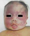
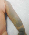
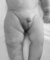
Sturge-Weber syndrome can be associated with facial port-wine stains and intracranial calcification, and concurrent Klippel-Trenaunay-Weber syndrome has been reported. Klippel-Trenaunay-Weber syndrome is a rare congenital mesodermal phakomatosis characterized by cutaneous hemangiomas, venous varicosities and soft tissue or bone hypertrophy of the affected extremities. This report is presented a rare case of the Sturge-Weber syndrome in combination with the Klippel-Trennaunay syndrome and phakomatosis pigmentovascularis in a 4-month-old infant. He showed nevus flameus on the right leg and both part of the face and back, leptomeningeal angiomatosis on right hemisphere, hypertrophy of the right leg, hemiconvulsion on the left and also evidences of congenital glaucoma and nevus of Ota. Very rare case combined with these three kinds of phakomatosis has been reported.
Figures


References
-
- Sturge WA. A case of partial epilepsy apparently due to a lesion of one of the vasomotor centers of the brain. Trans Clin Soc Lond. 1879;12:162–167.
-
- Barek L, Ledor S, Ledor K. The Klippel-Trenaunay syndrome: a case report and review of the literature. Mt Sinai J Med. 1982;49:66–70. - PubMed
-
- Deutsch J, Weissenbacher G, Widhalm K, Wolf G, Barsegar B. Combination of the syndrome of the Sturge-Weber and the syndrome of Klippel-Trenaunay. Klin Pediatr. 1976;188:464–471. - PubMed
-
- Stephan MJ, Hall BD, Smith DW, Cohen MM. Macrocephaly in association with unusual cutaneous angiomatosis. J Pediatr. 1975;87:353–359. - PubMed
Publication types
MeSH terms
LinkOut - more resources
Full Text Sources

