. 2006 Jan 11;25(1):244-54.
doi: 10.1038/sj.emboj.7600907. Epub 2005 Dec 15.

Huckebein-mediated autoregulation of Glide/Gcm triggers glia specification

Affiliations

Huckebein-mediated autoregulation of Glide/Gcm triggers glia specification

Rossana De Iaco et al. EMBO J. .

Abstract

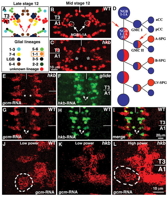
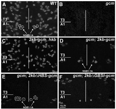
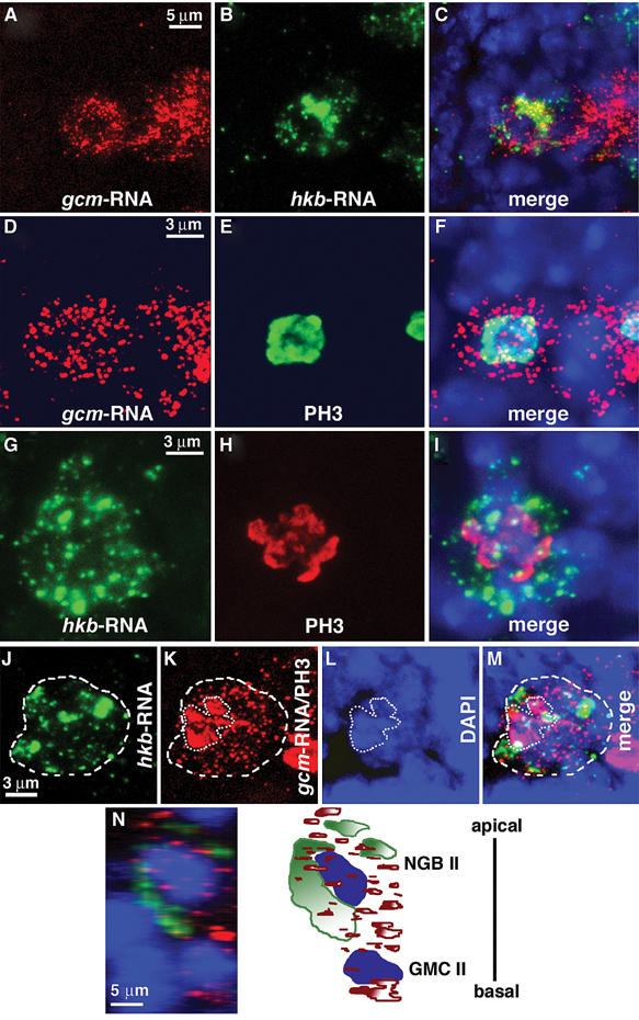
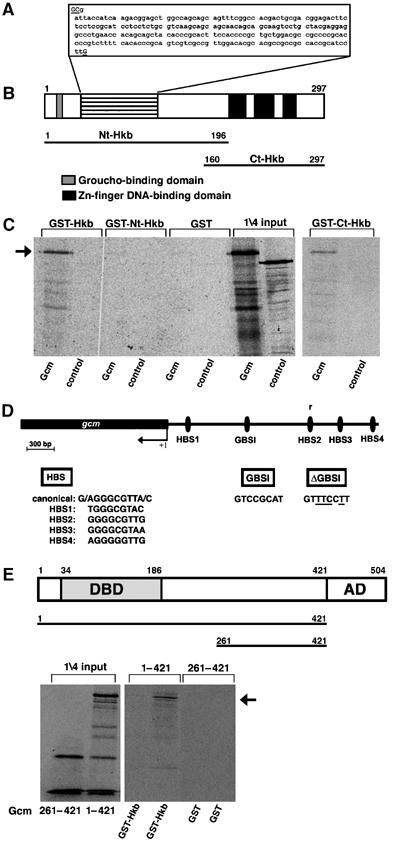
Cell specification in the nervous system requires patterning genes dictating spatio-temporal coordinates as well as fate determinants. In the case of neurons, which are controlled by the family of proneural transcription factors, binding specificity and patterned expression trigger both differentiation and specification. In contrast, a single gene, glide cell deficient/glial cell missing (glide/gcm), is sufficient for all fly lateral glial differentiation. How can different types of cells develop in the presence of a single fate determinant, that is, how do differentiation and specification pathways integrate and produce distinct glial populations is not known. By following an identified lineage, we here show that glia specification is triggered by high glide/gcm expression levels, mediated by cell-specific protein-protein interactions. Huckebein (Hkb), a lineage-specific factor, provides a molecular link between glide/gcm and positional cues. Importantly, Hkb does not activate transcription; rather, it physically interacts with Glide/Gcm thereby triggering its autoregulation. These data emphasize the importance of fate determinant cell-specific quantitative regulation in the establishment of cell diversity.


Figures

Figure 1
Figure 1
hkb controls glial differentiation in the NGB1-1A lineage. Unless otherwise specified, panels in this and in following figures show ventral views of the embryonic ventral cord; T3 and A1 indicate, respectively, third thoracic and first abdominal segments; anterior is to the top and the vertical line indicates the midline. (A) Schematic drawing of Repo labeled cells in T3 and A1 segments of a wild-type (WT)_ embryo at late stage 12. Glial subsets are identified by the expression of lineage-specific markers indicated by different colors. Symbols as in Ragone et al (2003). (B, C) Mid stage 12 embryos, WT (B) or hkb (C), labeled with glial-specific antibody anti-Repo. Arrows and dashed lines indicate NGB1-1A-derived glia. Asterisks in (C) indicate the absence of Repo labeling at the position normally taken by NGB1-1A-derived glia. (D) NGB1-1A lineage as proposed by Udolph et al (2001). Glial and neuronal potentials/components are indicated by red and blue, respectively. The first division of NGB1-1A gives rise to a ganglion mother cell (GMC I) that produces neurons (aCC and pCC), whereas the second division (NGB1-1A (II)) gives rise to a GMC (GMC II) that produces one neuron (n) and one glial cell (A-SPG). B-SPG and LV-SPG arise from later divisions. Red arrows indicate the two cells in which gcm mRNA is detected at stage 11. (E–L) Late stage 11 embryos. (E) hkb embryo labeled with gcm riboprobe (asterisks indicate the position normally taken by gcm expressing cells in the NGB1-1A lineage). (F) gcm embryo labeled with hkb riboprobe. (G–I) WT embryo labeled with gcm (G) and hkb (H) riboprobes. (I) Merge of (G and H). Arrows in (F–I) indicate cells of the NGB1-1A lineage. (J–L) WT (J) or hkb (K, L) embryos labeled with gcm riboprobe. Note the very low levels of gcm expression in mutant (K) compared to WT (J, dashed line) NGB1-1A lineage. (L) Same embryo as in (K) analyzed at high photomultiplicator power to amplify signal. Scale bars: 10 μm in (B, C, J–L) and 20 μm in (E–I).
Figure 2
Figure 2
NGB1-1A-derived glial cells depend on 2kb-gcm promoter. Late stage 12 embryos labeled with anti-Repo. (A) NGB1-1A glia are shown in a wild type (WT) by arrows and dashed lines. (B) No Repo positive nuclei are present in a gcm embryo (gcm). (C) NGB1-1A glial cells are absent in hkb embryo carrying the 2kb-gcm transgene (see asterisks), but present in gcm embryo carrying the same (D) or 2kbΔHBS-gcm (E) transgenes (see arrows) (100% of the animals, n=15). (F) NGB1-1A glia are absent in gcm embryos carrying the 2kbΔGBSI-gcm transgene (40% of the animals, n=50), see asterisks. Note that several transgenes were analyzed for each construct. To take into account position effects, transgenes of comparable strength were used. Scale bar: 10 μm.
Figure 3
Figure 3
hkb and gcm are coexpressed in NGB1-1A (II). NG1-1A lineage multiple labelling in wild-type (WT) embryos. (A–C) Triple labeling: (A) gcm riboprobe, (B) hkb riboprobe, (C) is a merge showing DAPI (blue) nuclear labeling. (D–F) Triple labeling: (D) gcm riboprobe, (E) anti-PH3 mitotic marker, (F) is a merge showing DAPI. (G–I) Triple labeling: (G) hkb riboprobe, (H) anti-PH3, (I) is a merge showing DAPI. (J–M) quadruple labeling: (J) hkb riboprobe (dashed line), (K) anti-PH3/gcm riboprobe, (L) DAPI labeling (dotted line), (M) is a merge. In (K), the same secondary antibody was used to reveal gcm RNA (dashed line) and PH3 (dotted line) because primary antibodies used were raised in the same species, but subcellular localization of gcm RNA and PH3 enables to distinguish between the two stainings. (N) 90° rotation to show labeling along the Z-axis. Triple labeling at two cell stage: hkb riboprobe (green), gcm riboprobe (red) and DAPI (blue). hkb-RNA is localized only in NGB1-1A II, whereas gcm-RNA is localized both in NGB and in GMC II, as schematically represented in the right panel. Scale bars: 5 μm in (A–C and N) and 3 μm in (D–M).
Figure 4
Figure 4
Hkb predicted ORF and interaction with Gcm. (A) Nucleotide sequence in the region indicated by horizontal lines in (B). Underlined nucleotides are absent in the published sequence (Bronner et al, 1994). (B) Organization of the predicted Hkb ORF. Boxes indicate the Zn-finger motifs (black) and the Groucho binding domain (gray) (Goldstein et al, 1999). Lines in (B) below the ORF indicate the Hkb truncated forms used to map interaction domains in GST pull-down assays. (C) Autoradiography of a pull-down assay using full length Gcm (amino acids 1–504) and GST–Hkb derivatives immobilized on glutathione-agarose beads. Binding of full-length Hkb protein (GST–Hkb) and Hkb C-terminal part (GST–Ct-Hkb) is indicated by an arrow. Luciferase (Luc) in vitro translated protein is used as a control. 1/4 of the input is shown in the right part of the panel. (D) Four Hkb binding sites (HBS1–4) are present in the 2kb-gcm promoter, one of which is in the opposite orientation (r). Canonical HBS represents the site identified by Kuhnlein et al (1997). GBSI indicates the Gcm binding site present in the 2kb-gcm promoter (Ragone et al, 2003). +1 indicates the transcription start site. (E) Gcm ORF: DBD indicates DNA binding domain, and AD, activation domain (see for a review Van De Bor and Giangrande, 2002). Lines below ORF indicate the in vitro translated products used to map interaction domains in GST pull-down assay shown in the bottom panel. Arrow indicates binding of GST–Hkb with translated Gcm 1-421 product. Note that GST pull-down assays entail a DNAse treatment, which eliminates possible DNA contamination.
Figure 5
Figure 5
Hkb binds to its target sequences but does not act as a transcription factor. (A) Gel-shift assay showing DNA binding of a purified GST-–Hkb fusion protein (Hkb). Labeled 27-mers corresponding to each of the four HBSs, to the canonical HBS (Kuhnlein et al, 1997) or to nonspecific DNA (NS: GCATGGACCAACATTGACACCGCTTTG) were used in the assay. Binding is indicated by arrows. (B) Hkb binding is abolished when HBS2 or HBS3 carrying point mutations are used (HBS2mut and HBS3mut, respectively, mutant nucleotides underlined). Binding to the canonical site is shown as a positive control. Arrow indicates the position of bound 27mers. (C) Competition gel-shift assay on canonical HBS and HBS1. S and NS indicate specific and nonspecific cold competitors, respectively (X indicates folds of excess, 0 indicates the absence of competitor). (D) pBLCAT5 reporter constructs containing either HBS1 or HBS2 (HBS1 and HBS2) were cotransfected with one (pPAC, pPAC-Gcm or pPAC-Hkb) or two (pPAC-Gcm and pPAC-Hkb) expression vectors. CAT assay data were normalized by using control reporter vector pBLCAT5. Each bar represents the average of at least three measurements, and error bars indicate standard error.
Figure 6
Figure 6
Gcm–Hkb synergistic activity. (A) Gel-shift assay showing DNA binding on a 30-mer containing GBSI. Arrow indicates binding of GST–Gcm N-terminal region (amino acids 1–261) to its target. Arrowhead indicates the additional band induced by incubation with both GST–Gcm N-terminal and GST–Hkb. Note that GST–Hkb does not, on its own, bind GBSI. Band shift induced by GST–Hkb is progressively removed by adding increasing amounts of anti-Hkb, but not by adding anti-Flag, used as nonspecific antibody (NS). Bracket indicates degraded GST–Gcm N-terminal products. (B) Cotransfection of reporter constructs containing either wild type (WT) or mutated GBSI (GBSI and ΔGBSI, see Figure 4) with pPAC-Gcm or pPAC-Hkb alone, or with pPAC-Gcm in combination with one of the Hkb-containing plasmids (pPAC-Hkb, pPAC-Ct-Hkb or pPAC-Nt-Hkb). (C) Cotransfection assays as in (B) but using reporter constructs containing either WT or mutated GBS1, a GBS that is three times more active than GBSI (Ragone et al, 2003; GBSC in Miller et al, 1998). CAT values obtained upon cotransfection with pPAC-Gcm and WT reporter were arbitrarily given a value of 1 and used for normalization. Each bar represents the average of at least three measurements, and error bars indicate standard error.
Figure 7
Figure 7
Role of Hkb and Gcm in NGB1-1A specification. (A–E″) Ventral views of stage 16 embryonic ventral cord carrying the P101 SPG marker; T3 and A1 indicate, respectively, third thoracic and first abdominal segments; anterior to the top and vertical line indicates the midline. β-gal (SPG), Repo double labeling. Left panels show β-gal, mid panels, Repo and right panels, merges. In all overexpression experiments, sca-GAL4 was used as a driver. (A–A″) labeling in wild-type (WT) embryo. Square brackets indicate A- and B-SPG (note that LV-SPG cannot be seen in this focal plane), arrowhead in (A) indicates lateral SPG, a cell that flanks A- and B-SPG cells, but is not derived from NGB1-1A lineage. (B–B″) labeling upon hkb overexpression induces additional SPG (B) and Repo (B′) labeling, close to the position at which A- and B-SPG are normally present. (C–C″) labeling upon combined gcm and hkb overexpression. Note the presence of additional SPG labeling in thoracic and abdominal segments (thick arrow). (D–D″) labeling upon gcm expression. (E–E″) labeling upon gcm expression in hkb embryo. (F–G″) Ventral views of stage 16 ventral cord of rA87/+ embryos. β-gal (rA87), Repo double labeling as above. (F–F″) labeling in a WT rA87/+ embryo. (G–G″) labeling in a sca-GAL4/rA87; UAS-hkb/+ embryo. Square brackets in (F and F″) indicate β-gal/Repo positive cells at the position of A- and B-SPG. Colocalization of additional Repo and β-gal labeling is indicated by arrows. Scale bar: 10 μm.
Figure 8
Figure 8
Hkb induces gcm expression in vivo. (A) gcm mRNA is expressed in one cell per hemisegment at stage 11 in a wild-type (WT) embryo (asterisks). (B) Several gcm expressing cells, indicated by white asterisks, are detectable in embryos overexpressing hkb (UAS-Hkb). sca-GAL4 driver was used for overexpression. Abdominal segments 7 and 8 (A7 and A8) are shown in (A) and (B). gcm expression profile at stage 14 in WT (C) and UAS-hkb (D) embryos. Thick arrows indicate persistent gcm expression in hkb overexpressing embryos. Scale bar: 10 μm.
Figure 9
Figure 9
Glial differentiation requires gcm autoregulation. Ventral views of stage 16 embryos, Repo labeling upon gcm overexpression (sca-GAL4) in wild type (WT) (A) or gcm background (B). Symbols as in Figure 1. Scale bar: 10 μm.

Similar articles

Cited by

References

    1. Akiyama-Oda Y, Hosoya T, Hotta Y (1998) Alteration of cell fate by ectopic expression of Drosophila glial cells missing in non-neural cells. Dev Genes Evol 208: 578–585 - PubMed
    1. Akiyama-Oda Y, Hosoya T, Hotta Y (1999) Asymmetric cell division of thoracic neuroblast 6–4 to bifurcate glial and neuronal lineage in Drosophila. Development 126: 1967–1974 - PubMed
    1. Akiyama Y, Hosoya T, Poole AM, Hotta Y (1996) The gcm-motif: a novel DNA-binding motif conserved in Drosophila and mammals. Proc Natl Acad Sci USA 93: 14912–14916 - PMC - PubMed
    1. Allan DW, Park D, St Pierre SE, Taghert PH, Thor S (2005) Regulators acting in combinatorial codes also act independently in single differentiating neurons. Neuron 45: 689–700 - PubMed
    1. Allan DW, Thor S (2003) Together at last: bHLH and LIM-HD regulators cooperate to specify motor neurons. Neuron 38: 675–677 - PubMed

Publication types