. 2005 Dec 21;24(24):4224-36.
doi: 10.1038/sj.emboj.7600888. Epub 2005 Dec 15.

A 'Collagen Hug' model for Staphylococcus aureus CNA binding to collagen

Affiliations

A 'Collagen Hug' model for Staphylococcus aureus CNA binding to collagen

Yinong Zong et al. EMBO J. .

Erratum in

  • EMBO J. 2006 Feb 22;25(4):921

Abstract

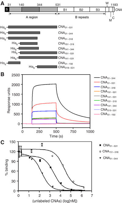
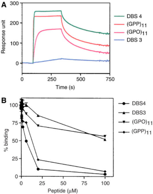
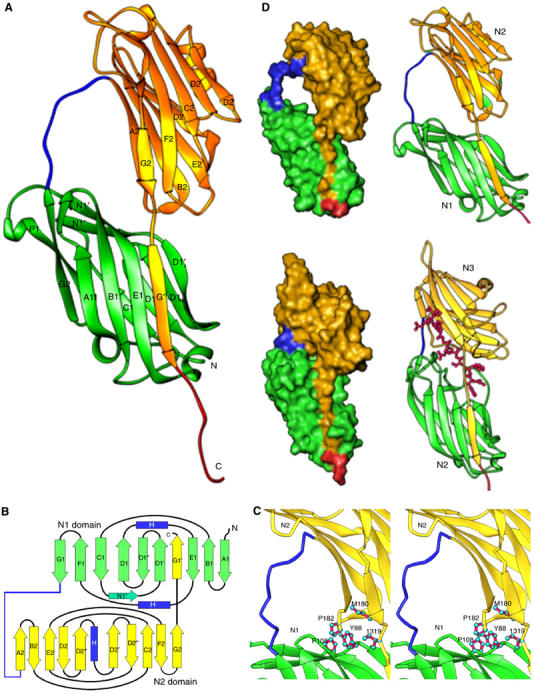
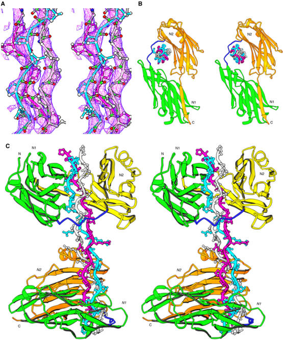
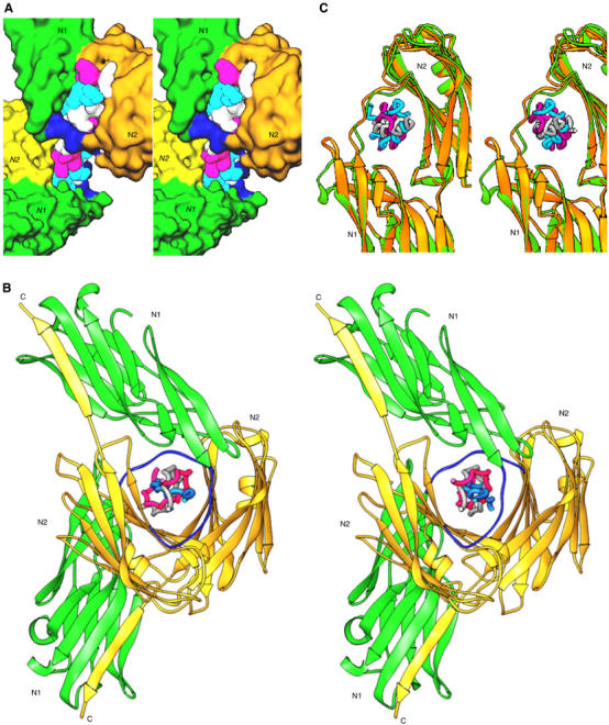
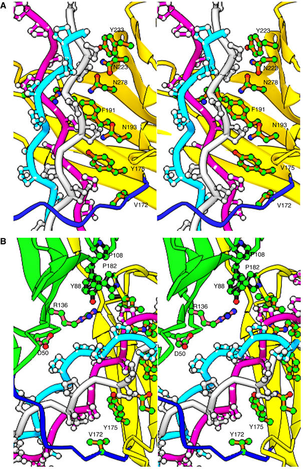
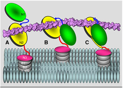
The structural basis for the association of eukaryotic and prokaryotic protein receptors and their triple-helical collagen ligand remains poorly understood. Here, we present the crystal structures of a high affinity subsegment of the Staphylococcus aureus collagen-binding CNA as an apo-protein and in complex with a synthetic collagen-like triple helical peptide. The apo-protein structure is composed of two subdomains (N1 and N2), each adopting a variant IgG-fold, and a long linker that connects N1 and N2. The structure is stabilized by hydrophobic inter-domain interactions and by the N2 C-terminal extension that complements a beta-sheet on N1. In the ligand complex, the collagen-like peptide penetrates through a spherical hole formed by the two subdomains and the N1-N2 linker. Based on these two structures we propose a dynamic, multistep binding model, called the 'Collagen Hug' that is uniquely designed to allow multidomain collagen binding proteins to bind their extended rope-like ligand.


Figures

Figure 1
Figure 1
(A) The domain organization of S. aureus CNA and different constructs. The collagen binding A region is followed by B repeats. S, signal peptide; W, cell wall anchoring region containing the LPETG motif; M, transmembrane segment; and C, cytoplasmic tail. The three subdomains of A-region are from residues 31–140 (N1), 141–344 (N2), and 345–531 (N3). The previously identified minimum collagen-binding domain is from residues 151–318. CNA fragments constructed as N-terminal His-tag fusion proteins are illustrated. (B) Representative Biacore sensorgrams of different CNA fragments passed over collagen. The same concentration of purified CNA fragments (10 μM) was passed over a bovine type I collagen-coated surface. Injection started at ∼140 s and ended at ∼550 s. Responses from a blank surface were subtracted from the responses from the collagen-coated surface. (C) Inhibition of the binding of CNA31–344 to type I collagen by different CNA fragments. Biotin-labeled CNA31–344 (100 nM) was mixed with increasing concentrations of unlabeled CNA31-344 (inverted triangles), CNA31–531 (squares), and CNA151–318 (open circles), and then incubated in wells coated with bovine type I collagen.
Figure 2
Figure 2
(A) Representative sensorgrams of the synthetic collagen peptides passed over CNA31–344. The same concentrations of four collagen peptides (1 μM) were passed over a CNA31–344-coated surface. Injection started at ∼95 s and ended at ∼335 s. Responses from a blank surface were subtracted from responses from collagen-coated surface. (B) Inhibition of the binding of CNA31–344 to type I collagen by synthetic collagen peptides. CNA31–344 (10 nM) was preincubated with increasing amounts of synthetic collagen peptides DBS4 (•), DBS3 (▴), (GPO)11 (▾), and (GPP)11 (⧫), and then added to wells coated with bovine type I collagen.
Figure 3
Figure 3
Crystal structure of the apo-CNA31–344. (A) Apo-CNA31–-344 consists two domains, N1 (Green) and N2 (Yellow). Residues 164–173 form a linker (blue in color) that joins the N1 and N2 domains. (B) Cartoon representation of the apo-CNA31–344 crystal structure. Strands are represented as arrows (green for the N1 domain and yellow for the N2 domain), and helices in blue color. The linker joining the N1 and N2 domains is represented in blue and all other loop regions in black. Images of the molecular structures were prepared using RIBBONS program (Carson, 1997). (C) The inter domain hole in apo-CNA31–344 is formed by the linker that connects N1 and N2 domains and hydrophobic residues contributed by both N1 and N2 domains. (D) Comparison of apo-CNA31–344 and SdrGN2N3 crystal structures. The crystal structure of apo-CNA31–344 (top) is compared to the ligand bound SdrGN2N3 crystal structure (bottom) to illustrate the common C-terminal extension ‘donor strand' mode and the length differences in the inter domain linker region (blue).
Figure 4
Figure 4
The crystal structure of (CNA31–344)2–collagen-like peptide complex. (A) The representative simulated annealing omit map for the collagen-like peptide is viewed at 1.5σ. (B) The triple helical collagen peptide is seen parked in the hole between N1 and N2 domains. The three chains of the collagen peptide are named as leading (magenta), middle (cyan) and trailing (white) as viewed from their N-termini. (C) Two CNA31–344 molecules interact with one collagen peptide in an identical manner. The leading and trailing chains peptide interact with N2 domain β sheet, and the middle chain with N1. The N1–N2 linker covers the leading and trailing chains, and holds the rope like ligand in place.
Figure 5
Figure 5
(A) A surface plot of the collagen peptide with the two CNA31–344 molecules. The N1–N2 linker regions are shown in blue. (B) Two CNA31–344 molecules in one complex molecule have a rotational difference of 120° when viewed down the collagen peptide from N-terminus. (C) The conformational switch of a loop region (residue 138–148) between apo-CNA31–344(green) and ligand bound CNA31–344 (yellow) for stabilizing the bound ligand.
Figure 6
Figure 6
Specific interactions between CNA31–344 and the collagen peptide ligand. (A) The triple helical collagen peptide mainly interacts with residues in the trench of the N2 domain (yellow). Stacking hydrophobic interactions between the proline residues of the ligand and the bulky hydrophobic Tyr and Phe residues of the N2 domain are observed. Val172 from the N1–N2 linker (blue) also participates in such hydrophobic interactions. (B) The minimal ligand interactions contributed by the N1 domain (green) are presented. The hydrophobic inter-domain ‘locking' arrangement can be seen between residues Pro108 and Tyr88 of N1 domain, and Pro182 and Met180 from N2 domain, and the Ile319 from the C-terminal extension.
Figure 7
Figure 7
A hypothetical ‘Collagen Hug' model shown in a cartoon representation. (A) Collagen triple helix is initially associated with the N2 domain. (B) The collagen is then wrapped by the N1–N2 linker and the N1 domain. (C) The N1 domain interacts with the N2 domain via multiple hydrophobic interactions and finally the C-terminal latch is introduced in the N1 domain to secure the ligand in place.

References

    1. Bella J, Eaton M, Brodsky B, Berman HM (1994) Crystal and molecular structure of a collagen-like peptide at 1.9 Å resolution [see comment]. Science 266: 75–81 - PubMed
    1. Brodsky B, Persikov AV (2005) Molecular structure of the collagen triple helix. Adv Protein Chem 70: 301–339 - PubMed
    1. Brunger AT, Adams PD, Clore GM, DeLano WL, Gros P, Grosse-Kunstleve RW, Jiang JS, Kuszewski J, Nilges M, Pannu NS, Read RJ, Rice LM, Simonson T, Warren G (1998) Crystallography & NMR system: a new software suite for macromolecular structure determination. Acta Crystallogr D Biol Crystallogr 54 (Part 5): 905–921 - PubMed
    1. Carson M (1997) Ribbons. Methods Enzymol 277: 493–505 - PubMed
    1. Clemetson KJ, Clemetson JM (2001) Platelet collagen receptors. Thromb Haemost 86: 189–197 - PubMed

Publication types

MeSH terms