. 2005 Dec 21;24(24):4291-303.
doi: 10.1038/sj.emboj.7600883. Epub 2005 Dec 15.

Interplay between 7SK snRNA and oppositely charged regions in HEXIM1 direct the inhibition of P-TEFb

Affiliations

Interplay between 7SK snRNA and oppositely charged regions in HEXIM1 direct the inhibition of P-TEFb

Matjaz Barboric et al. EMBO J. .

Abstract

Transcription elongation of eukaryotic genes by RNA polymerase II depends on the positive transcription elongation factor b (P-TEFb). When sequestered into the large complex, P-TEFb kinase activity is inhibited by the coordinate actions of 7SK small nuclear RNA (7SK snRNA) and hexamethylene bisacetamide (HMBA)-induced protein 1 (HEXIM1). We found that the basic region in HEXIM1 directs its nuclear import via two monopartite and two bipartite nuclear localization sequences. Moreover, the arginine-rich motif within it is essential for its binding to 7SK snRNA, P-TEFb, and inhibition of transcription. Notably, the basic region interacts with the adjacent acidic regions in the absence of RNA. The removal of the positive or negative charges from these regions in HEXIM1 leads to its sequestration into the large complex and inhibition of transcription independently of the arginine-rich motif. Finally, the removal of the negative charges from HEXIM1 results in its subnuclear localization into nuclear speckles. We propose a model where the interplay between 7SK snRNA and oppositely charged regions in HEXIM1 direct its binding to P-TEFb and subcellular localization that culminates in the inhibition of transcription.


Figures

Figure 1
Figure 1
BR in HEXIM1 contains multiple predicted NLSs. (A) HEXIM1 protein is presented as a white rectangle. Black boxes represent two basic (BR; BR1 and BR2) and two acidic (AR; AR1 and AR2) regions and the encircled signs above them represent the charge of these regions. The sequence numberings correspond to the N-terminus, the boundaries of the BRs and ARs, and the C-terminus, respectively. (B) Alignment of the BRs of human, mouse, chicken, zebrafish, and fish HEXIM1 and HEXIM2 proteins. Reverse type indicates basic amino-acid identity whereas shaded boxes indicate basic amino-acid similarity. (C) Predicted monopartite and bipartite NLSs within the BR in HEXIM1 is presented. (D) HEXIM1 proteins used in the first part of the study. Hex1 represents the primary amino-acid sequence of the BR in HEXIM1, which was a subject to site-directed mutagenesis. Numbers above this sequence indicate a disruption of the corresponding basic cluster. The names of wild-type and mutant Hex1 proteins are presented on the right-hand side of the panel. A capital letter B symbolizes basic cluster, whereas small letter m symbolizes the replacement of corresponding basic clusters with alanines.
Figure 2
Figure 2
Four distinct NLSs direct nuclear import of HEXIM1. (A, B) f.Hex1 proteins (green) that were expressed in HeLa cells are indicated above and below the microscopic images. Cell nuclei were visualized by propodium iodide (PI; red). The images depict the merge of the f.Hex1 and PI images. (C) HeLa cell lysates, which expressed f.Hex1 or f.Hex1mB1256 from corresponding plasmid effectors (10 μg), were incubated with h6.x.Impα as indicated. Arrows to the left indicate bound f.Hex1 proteins (pd) and 10% inputs (i) of the proteins used in the assay, respectively. (D) YFP (image 1) and Hex1.YFP (images 2 and 3) proteins (green) were expressed in HeLa cells. The panel marked Hex1.YFP/C23 depict the merge of the Hex1.YFP (green) and C23 (red) images. (E) BR.YFP proteins (green) that were expressed in HeLa cells are indicated above the microscopic images. Where indicated, the images depict the merge of the respective BR.YFP (green) and C23 (red) images. Lower parts of the panels D and E indicate the cell nuclei of which the DNA was counterstained by DAPI (blue).
Figure 3
Figure 3
The disruption of the ARM in HEXIM1 disables its binding to 7SK snRNA in vitro and inhibition of transcription and binding to P-TEFb in vivo. (A) Chimeric GST.Hex1 proteins are indicated above the autoradiograph. α-32P-labeled 7SK snRNA was present in all reactions. Arrow to the left indicates the free 7SK snRNA probe and the presence of 7SK snRNA:GST.Hex1 RNA–protein complexes is bracketed. (B) HeLa cells expressed plasmid reporter pG6TAR (0.4 μg; bars 1–6). Proteins that were coexpressed from corresponding plasmid effectors (Gal4.CycT1, 0.6 μg; f.Hex1, 0.8 μg) with the plasmid reporter are presented below CAT data. The lower panels present the expression of f.Hex1 proteins as indicated by the arrow. (C) f.Hex1 proteins that were expressed in HeLa cells from corresponding plasmid effectors (10 μg; lanes 2–6) and immunoprecipitated by anti-FLAG M2 beads are indicated above the Western blots. Arrows to the left indicate the bound P-TEFb and the amounts of immunoprecipitated f.Hex1 proteins, respectively.
Figure 4
Figure 4
The interaction between the BR and AR within HEXIM1 in vitro is abolished in the presence of RNA. (A) The mutant GST.Hex1 and h6.x.Hex1 proteins are presented as white rectangles. Black boxes depict BR and AR as in Figure 1A. The numberings above the schematic correspond to the N- and/or C-terminal boundaries of the proteins. The names of the proteins are presented on the left-hand side of the panel. (B, C) Chimeric GST.Hex1 proteins were incubated with the mutant h6.x.Hex1 protein in the presence or absence of RNase A as indicated above the Western blot. Arrow to the left in panel B indicates the bound h6.x.Hex1 protein. In the panel C, the h6.x.Hex1 proteins are bracketed. Left part of the panel (lanes 1–6) represents the bound h6.x.Hex1 proteins, whereas the right part of the panel (lanes 7–9) represents the 20% inputs of the proteins used in the assay. Numbers to the right indicate relative molecular mass markers (in kDa).
Figure 5
Figure 5
The disruption of the BR in HEXIM1 enables the ARM-independent inhibition of transcription and binding to P-TEFb in vivo. (A) HeLa cells expressed plasmid reporter pG6TAR (0.4 μg; bars 1–6). Proteins that were coexpressed from corresponding plasmid effectors (Gal4.CycT1, 0.6 μg; f.Hex1, 0.8 μg) with the plasmid reporter are presented below CAT data. The lower panel presents the expression of f.Hex1 proteins as indicated by the arrow. (B) f.Hex1 proteins that were expressed in HeLa cells from corresponding plasmid effectors (1 μg) are indicated above and below the microscopic images. f.Hex1 proteins (green) and nuclei (red) were visualized by laser confocal microscopy. (C) f.Hex1 proteins that were expressed in HeLa cells from corresponding plasmid effectors (10 μg; lanes 1–3) and immunoprecipitated by anti-FLAG M2 beads are indicated above the Western blots. Arrows to the left indicate bound P-TEFb and the amounts of immunoprecipitated f.Hex1 proteins, respectively. (D) Total cell lysates of untransfected HeLa cells and those that expressed the indicated f.Hex1 proteins were subjected to glycerol gradient (10–30%) sedimentation analysis. The lysates were mock or RNase A treated as indicated above the Western blots. Arrows to the right indicate the presence of endogenous CycT1 and HEXIM1 (Hex1) proteins, and f.Hex1 proteins that were expressed from corresponding plasmid effectors (6 μg). Numberings below the Western blots correspond to particular fractions obtained from the sedimentation analysis. SC and LC indicate fractions containing the small and large complexes, respectively.
Figure 6
Figure 6
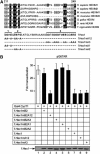
The disruption of the AR1 in HEXIM1 enables the ARM-independent inhibition of transcription. (A) Alignment of human, mouse, chicken, zebrafish, and fish HEXIM1 and HEXIM2 proteins and f.Hex1 proteins used in the study. The sequence numberings correspond to the boundaries of the ARs in HEXIM1. Reverse type indicates acidic amino-acid identity, whereas shaded boxes indicate acidic amino-acid similarity. The lower panel represents the primary amino-acid sequence of the AR in HEXIM1, which was a subject to site-directed mutagenesis. Numbers above this sequence indicate a disruption of the corresponding acidic cluster. The names of Hex1 proteins are presented on the right-hand side of the panel. A capital letter A symbolizes acidic cluster, whereas a small letter m symbolizes the replacement of corresponding acidic clusters with alanines. (B) HeLa cells expressed plasmid reporter pG6TAR (0.4 μg; bars 1–9). Proteins that were coexpressed from corresponding plasmid effectors (Gal4.CycT1, 0.6 μg; f.Hex1, 0.8 μg) with the plasmid reporter are presented below CAT data. The lower panel presents the expression of f.Hex1 proteins as indicated by the arrow.
Figure 7
Figure 7
The disruption of the AR in HEXIM1 enables the ARM-independent binding to P-TEFb. (A) Chimeric GST.Hex1 proteins that were used in EMSAs are indicated above the autoradiograph. α-32P-labeled 7SK snRNA was present in all reactions. Arrows to the left indicate the free 7SK snRNA probe and the presence of 7SK snRNA:GST.Hex1 RNA–protein complexes. (B) HeLa cell lysates, which were treated with ActD, were incubated with the chimeric GST.Hex1 proteins as indicated. Arrows to the left indicate bound GST.Hex1 proteins (pd) and 20% inputs (i) of the proteins used in the assay, respectively. (C) Total cell lysates of HeLa cells that expressed the indicated f.Hex1 proteins were subjected to glycerol gradient (10–30%) sedimentation analysis. The lysates were mock or RNase A treated as indicated above the Western blots. Arrows to the right indicate the presence of f.Hex1 proteins that were expressed from corresponding plasmid effectors (6 μg). Numberings below the Western blots correspond to particular fractions obtained from the sedimentation analysis. SC and LC indicate fractions containing the small and large complexes, respectively.
Figure 8
Figure 8
The disruption of the AR1 in HEXIM1 leads to its localization into nuclear speckles. (A) Hex1.YFP proteins that were expressed in HeLa cells are indicated above the microscopic images. (B) HeLa cells expressed Hex1mB2A12.YFP (green) (images 1–9). The panels marked with SC-35, PML, and C23 (red) represent the images of nuclear speckles, PML nuclear bodies, and nucleoli, whereas the panels marked with merge depict the merge of the Hex1mB2A12.YFP and SC-35, PML, and C23 images, respectively. (C) Hex1.YFP proteins (green) that were expressed in HeLa cells are indicated above the microscopic images. The panel marked Hex1(1–286)mB2A12.YFP/C23 depict the merge of the Hex1(1-286)mB2A12.YFP (green) and C23 (red) images. (D) HeLa cells expressed plasmid reporter pG6TAR (0.4 μg; bars 1–5). Proteins that were coexpressed from corresponding plasmid effectors (Gal4.CycT1, 0.6 μg; f.Hex1, 0.8 μg) with the plasmid reporter are presented below CAT data. The lower panel presents the expression of f.Hex1 proteins as indicated by the bracket.

References

    1. Barboric M, Peterlin BM (2005) A new paradigm in eukaryotic biology: HIV Tat and the control of transcriptional elongation. PLoS Biol 3: e76. - PMC - PubMed
    1. Blazek D, Barboric M, Kohoutek J, Oven I, Peterlin BM (2005) Oligomerization of HEXIMI via 7SK snRNA and coiled-coil region directs the inhibition of P-TEFb. Nucleic Acids Res (in press) - PMC - PubMed
    1. Byers SA, Price JP, Cooper JJ, Li Q, Price DH (2005) HEXIM2, a HEXIM1 related protein, regulates P-TEFb through association with 7SK. J Biol Chem 280: 16360–16367 - PubMed
    1. Carmo-Fonseca M (2002) The contribution of nuclear compartmentalization to gene regulation. Cell 108: 513–521 - PubMed
    1. Chao SH, Price DH (2001) Flavopiridol inactivates P-TEFb and blocks most RNA polymerase II transcription in vivo. J Biol Chem 276: 31793–31799 - PubMed

Publication types

MeSH terms