Review
. 2005 Dec;1(4):e37.
doi: 10.1371/journal.ppat.0010037. Epub 2005 Dec 16.

Invasion by P. falciparum merozoites suggests a hierarchy of molecular interactions

Affiliations
Review

Invasion by P. falciparum merozoites suggests a hierarchy of molecular interactions

Jake Baum et al. PLoS Pathog. 2005 Dec.

Abstract

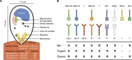
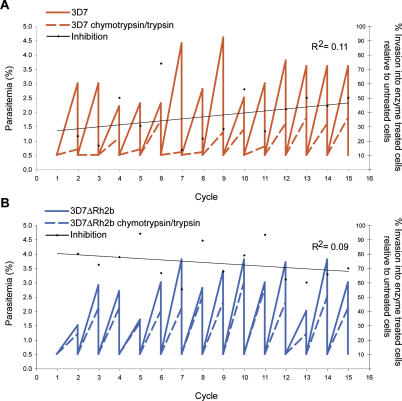
Central to the pathology of malaria disease are the repeated cycles of parasite invasion and destruction of human erythrocytes. In Plasmodium falciparum, the most virulent species causing malaria, erythrocyte invasion involves several specific receptor-ligand interactions that direct the pathway used to invade the host cell, with parasites varying in their dependency on these different pathways. Gene disruption of a key invasion ligand in the 3D7 parasite strain, the P. falciparum reticulocyte binding-like homolog 2b (PfRh2b), resulted in the parasite invading via a novel pathway. Here, we show results that suggest the molecular basis for this novel pathway is not due to a molecular switch but is instead mediated by the redeployment of machinery already present in the parent parasite but masked by the dominant role of PfRh2b. This would suggest that interactions directing invasion are organized hierarchically, where silencing of dominant invasion ligands reveal underlying alternative pathways. This provides wild parasites with the ability to adapt to immune-mediated selection or polymorphism in erythrocyte receptors and has implications for the use of invasion-related molecules in candidate vaccines.


Conflict of interest statement

Competing interests. The authors have declared that no competing interests exist.

Figures

Figure 1
Figure 1. A Number of Receptor–Ligand Interactions Mediate Merozoite Invasion of the Erythrocyte
(A) Estimates for the dimensions of the interaction between the apical merozoite tip and the erythrocyte surface suggest a finite number of receptors may, together with parasite ligands, direct invasion (dimensions based on [55]; receptor figures based on [56]). (B) Known receptor–ligand interactions between the invading merozoite and erythrocyte surface receptors and their enzyme sensitivities to neuraminidase (Nm), trypsin, and chymotrypsin (Chymo). S indicates sensitivity of invasion to this enzyme (inhibits invasion/ligand-binding). R indicates resistance (does not inhibit invasion/ligand-binding). Unknown ligands or receptors are indicated by a “?,” with unknown receptors E, X, Y, and Z known only from their enzyme sensitivities. Dots in table indicate that no data are available. Data are taken from [7,8,10–12,14,17,27,33,45].
Figure 2
Figure 2. Comparison of Transcription of Key Invasion Genes in 3D7, 3D7ΔRh2b, and D10
(A and B). Expression levels of EBA and PfRh genes controlled by the relative expression of either actin and histone 2b (two constitutively expressed genes) or msp2 (a gene expressed later on in the life cycle). Error bars represent the 95% confidence interval (CI) of values from two to three independent amplifications. (C and D) Affymetrix microarray comparison of log gene expression intensity (as measured by MOID) between cRNA isolated from late-schizont 3D7 and (C) 3D7ΔRh2b or (D) D10 parasites. Data shown are restricted to 548 genes whose expression profile matches clusters 4, 13, 14, 15 [34], including genes that encode proteins known to be involved in invasion. Color represents significance of the change in the level of gene expression as measured by the t-statistic (<2 black, >2 red). The control genes PfRh2b (knocked out) and EBA-140 (deleted in D10) and upregulation of PfRh3 are shown.
Figure 3
Figure 3. Targeted Disruption of PfRh3 Shows It Is Not Essential for the 3D7ΔRh2b Chymotrypsin-Resistant Pathway
(A) Disruption of the PfRh3 gene in 3D7. The pCC4-Rh3 plasmid contains the blasticidin-S deaminase selectable marker, a negative selectable marker (A. G. Maier and A. F. Cowman, unpublished data), and 5′ and 3′ Rh3 regions. PfRh3 is shown with homologous target sequences (shaded regions). The double-crossover integration events are shown for 3D7, resulting in the deletion of a 5′ region of the gene. Restriction enzymes are C (ClaI) and X (XbaI), with fragment sizes shown for a C/X digestion. RT-PCR amplification target is shown as a black bar. (B) Southern blot of genomic DNA from parasites shown digested with ClaI and XbaI and probed with the 5′ Rh3 flank from the pCC4-Rh3 vector. (C) RT-PCR of PfRh2a and PfRh3 from 3D7 and three knockout lines. (D) Invasion into enzyme-treated erythrocytes expressed as a percentage of that into untreated erythrocytes for 3D7 and three knockout lines. Values above each column indicate the mean % invasion, with error bars representing the 95% CI from three independent assays.
Figure 4
Figure 4. Changes in Protein Levels Do Not Underlie the 3D7ΔRh2b Chymotrypsin-Resistant Pathway
(A) Western blot of parasite culture supernatant material probed with rabbit polyclonal antibodies against the functional EBA and PfRh proteins. SERA5 is used as a loading control. Antibodies marked with an asterisk (*) indicate the same gel stripped and reprobed with a different antibody. (B) Invasion into chymotrypsin-treated erythrocytes for five parasite lines represented as the percentage of invasion to that into untreated erythrocytes. (C) Invasion into either untreated or chymotrypsin-treated erythrocytes in the presence of protein-G purified polyclonal rabbit antiserum raised against recombinant EBA-140 and EBA-175 (or both together). Values are represented as the percentage of invasion in the presence of NRS. Error bars represent the 95% CI from three independent assays.
Figure 5
Figure 5. Long-Term Selection of (A) 3D7 and (B) 3D7ΔRh2b in Erythrocytes Pretreated with Chymotrypsin (1.5 mg/ml) and Trypsin (0.1 mg/ml) Shows No Significant Change in Receptor Dependency
Parasitemia was measured by microscopy; then cultures were diluted down to 0.5% parasitemia at 48-h intervals. Cultures were maintained for greater than 30 d. Secondary y-axis represents invasion into enzyme-treated erythrocytes as a percentage of invasion into untreated erythrocytes, with a regression line plotted across the values.

References

    1. Miller LH, Baruch DI, Marsh K, Doumbo OK. The pathogenic basis of malaria. Nature. 2002;415:673–679. - PubMed
    1. Gaur D, Mayer DC, Miller LH. Parasite ligand-host receptor interactions during invasion of erythrocytes by Plasmodium merozoites. Int J Parasitol. 2004;34:1413–1429. - PubMed
    1. Friedman MJ, Blankenberg T, Sensabaugh G, Tenforde TS. Recognition and invasion of human erythrocytes by malarial parasites: Contribution of sialoglycoproteins to attachment and host specificity. J Cell Biol. 1984;98:1672–1677. - PMC - PubMed
    1. Mayer DC, Kaneko O, Hudson-Taylor DE, Reid ME, Miller LH. Characterization of a Plasmodium falciparum erythrocyte-binding protein paralogous to EBA-175. Proc Natl Acad Sci U S A. 2001;98:5222–5227. - PMC - PubMed
    1. Miller LH, Haynes JD, McAuliffe FM, Shiroishi T, Durocher JR, et al. Evidence for differences in erythrocyte surface receptors for the malarial parasites, Plasmodium falciparum and Plasmodium knowlesi . J Exp Med. 1977;146:277–281. - PMC - PubMed

Publication types

MeSH terms

Substances