. 2006 Jan;136(1):21-6.
doi: 10.1093/jn/136.1.21.

Copper transport protein (Ctr1) levels in mice are tissue specific and dependent on copper status

Affiliations

Copper transport protein (Ctr1) levels in mice are tissue specific and dependent on copper status

Yien-Ming Kuo et al. J Nutr. 2006 Jan.

Abstract

Studies were conducted to determine distribution of the copper transporter, Ctr1, a transmembrane protein responsible for cellular copper uptake, in adult mice and in suckling mice nursed by either copper-adequate (Cu+) or copper-deficient (Cu-) dams. Western immunoblot analyses, using immunopurified antibody, detected monomeric (23 kDa) and oligomeric forms of Ctr1 in the membrane fraction of several mouse organs. Immunohistochemical analyses detected abundant Ctr1 protein in liver canaliculi; kidney cortex tubules; small intestinal enterocytes; the choroid plexus and capillaries of brain; intercalated disks of heart; mature spermatozoa; epithelium of mammary ducts; and the pigment epithelium, outer limiting membrane, and outer plexiform layer of the retina. Duodenal Ctr1 distribution was different in the adult compared with suckling mice; adult mice demonstrated strong intracellular staining of the enterocyte, whereas apical staining predominated in suckling mice. In Cu- mice at postnatal d 16 (P16), Ctr1 staining was augmented in kidney, duodenum, and choroid plexus, compared with Cu+ mice. Brain immunoblot data indicated that Ctr1 protein in membrane fractions of Cu- mice was 56% higher than Cu+ mice. Cu- mice had lower hemoglobin (56% of Cu+), and lower copper concentration (% of Cu+) in liver (15%), brain (26%), and kidney (65%). These results suggest that Ctr1 protein is expressed in multiple tissues and found in higher levels in selected organs after perinatal copper deficiency. Enhanced Ctr1 levels and redistribution might compensate in part for the decrease in copper supply. Mechanisms for the enhancement in Ctr1 staining remain to be established.


Figures

FIGURE 1
FIGURE 1
Characterization of Ctr1 by Western analysis (A) and tissue distribution of Ctr1 in an adult mouse (B). Kidney (K) membranes from an adult P120 male mouse were subjected to Western immunoblot protocols using 30 µg protein and 12% SDS-PAGE under reducing conditions and a rabbit anti-Ctr1 immunopurified antibody. Preabsorption with excess antigen peptide blocked 3 main Ctr1 specific bands (arrows) corresponding to peptides with mobilities compared with standards of 23, 46, and 70 kDa, representing monomer, dimer, and trimeric forms of Ctr1 (A). Membranes from another adult mouse were compared by a similar protocol and loading (B). All 3 Ctr1 bands identified for kidney (A) were present in kidney in this mouse as were bands for liver (L), heart (H), brain (B), and small intestine enterocytes (I), although staining intensity was organ specific.
FIGURE 2
FIGURE 2
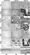
Immunohistochemical localization of Ctr1 in mouse liver, kidney, small intestine, brain, heart, testes, and retina at 10 wk of age. No staining was detected in the preimmune control serum (left column). Images in the middle and right columns were taken at 20 and 100× magnification, respectively. Ctr1 was localized in the canaliculi of the liver (arrow), in the cortex of the apical part of the kidney tubule epithelium (arrow), and cytoplasm of enterocytes (arrows). In the brain, Ctr1 was enriched in the choroid plexus epithelial cells lining the ventricular lumen (arrow). Ctr1 was localized in the intercalated discs of the heart (arrow). Ctr1 was highly expressed in the mature spermatozoa of the testes (arrow). In the retina, Ctr1 was detected in the retinal pigment epithelium, the outer limiting membrane, and the outer plexiform layer (arrows).
FIGURE 3
FIGURE 3
Immunohistochemical localization of Ctr1 in mouse mammary tissue. No staining was detected in the preimmune control serum (left column). Images in the middle and right columns were taken at and 100× magnification, respectively. Ctr1 was localized to the epithelium of the ducts and alveoli (arrow) of mammary glands taken from adult virgin, pregnant, lactating, and involuted (postlactating) mice.
FIGURE 4
FIGURE 4
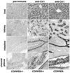
Immunohistochemical localization of Ctr1 in the liver, kidney, small intestine, and brain of copper-adequate and copper-deficient mice at P16. No staining was detected using the preimmune control serum (left column) for copper-adequate or copper-deficient tissues (data not shown). Data in the middle column are from a copper-adequate mouse and those in the right column from a copper-deficient mouse. Similar images from the additional mice were observed. Images were taken at 100× magnification. In the liver, we did not detect significant changes in Ctr1 expression upon copper depletion. In the kidney and duodenum, Ctr1 staining was more intense in samples from copper-deficient mice. In the brain, Ctr1 expression was also higher in the choroid plexus from copper-deficient mice.
FIGURE 5
FIGURE 5
The effect of copper deficiency on Ctr1 protein expression in suckling mouse brain. Four pairs of copper adequate (+) and copper deficient (−) male P18 mice, brothers of those used for immunocytochemistry, were used to evaluate Ctr1 expression in membrane fractions of whole brain. The mean density of both Ctr1 monomer and dimer bands in the Cu− lanes was 56% higher than Cu+ lanes in the membrane fraction immunoblot, P < 0.05. Each lane contained 25 µg protein. Ponceau S staining of the membrane indicated equivalent protein loading and transfer.

Similar articles

Cited by

References

    1. Prohaska JR, Gybina AA. Intracellular copper transport in mammals. J Nutr. 2004;134:1003–1006. - PubMed
    1. Vulpe CD, Packman S. Cellular copper transport. Annu Rev Nutr. 1995;15:293–322. - PubMed
    1. Petris MJ, Mercer JF, Culvenor JG, Lockhart P, Gleeson PA, Camakaris J. Ligand-regulated transport of the Menkes copper P-type ATPase efflux pump from the Golgi apparatus to the plasma membrane: a novel mechanism of regulated trafficking. EMBO J. 1996;15:6084–6095. - PMC - PubMed
    1. Dancis A, Haile D, Yuan DS, Klausner RD. The Saccharomyces cerevisiae copper transport protein (Ctr1p). Biochemical characterization, regulation by copper, and physiologic role in copper uptake. J Biol Chem. 1994;269:25660–25667. - PubMed
    1. Zhou B, Gitschier J. hCTR1: a human gene for copper uptake identified by complementation in yeast. Proc Natl Acad Sci U S A. 1997;94:7481–7486. - PMC - PubMed

Publication types