. 2005 Dec 19;171(6):1073-84.
doi: 10.1083/jcb.200503155.

Alpha4beta1-dependent adhesion strengthening under mechanical strain is regulated by paxillin association with the alpha4-cytoplasmic domain

Affiliations

Alpha4beta1-dependent adhesion strengthening under mechanical strain is regulated by paxillin association with the alpha4-cytoplasmic domain

Ronen Alon et al. J Cell Biol. .

Abstract

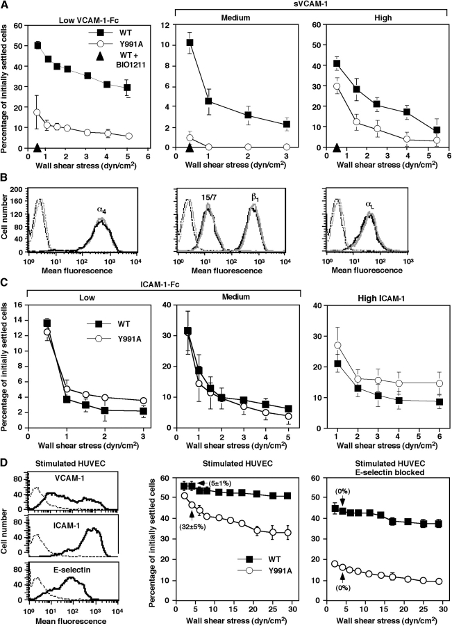
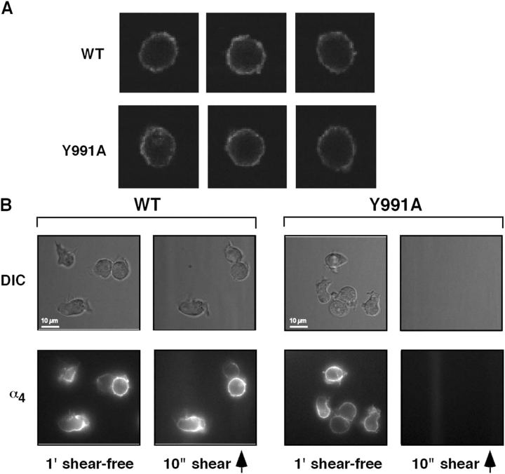
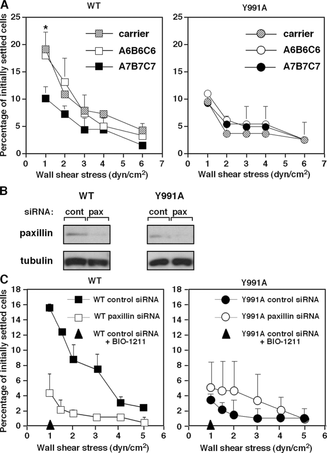
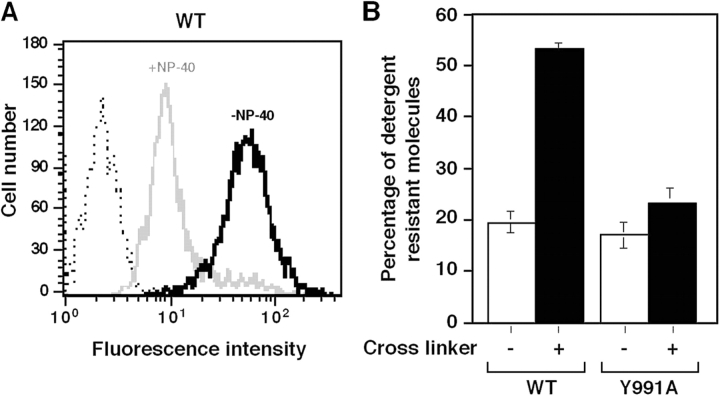
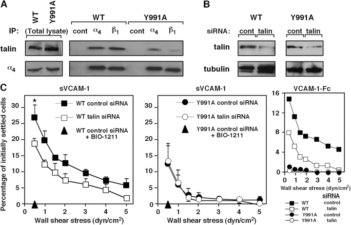
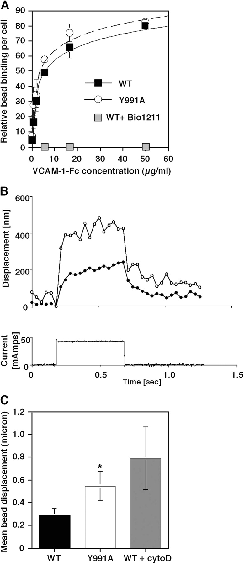
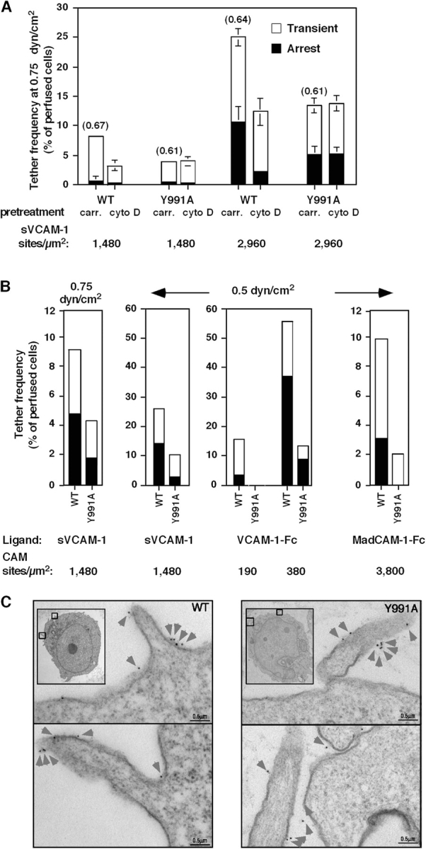
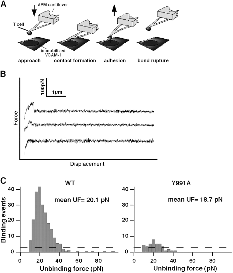
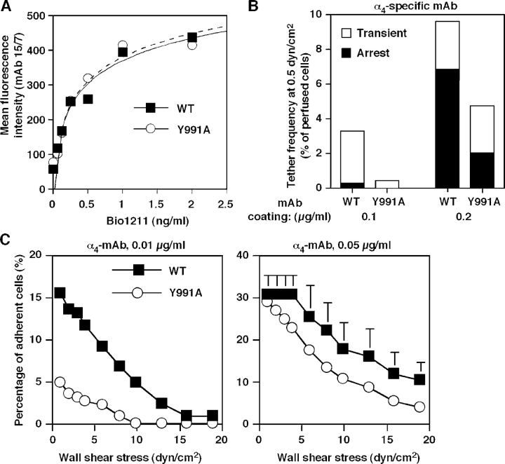
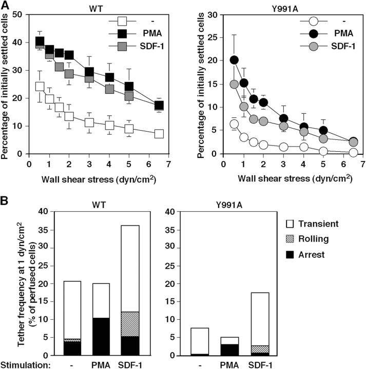
The capacity of integrins to mediate adhesiveness is modulated by their cytoplasmic associations. In this study, we describe a novel mechanism by which alpha4-integrin adhesiveness is regulated by the cytoskeletal adaptor paxillin. A mutation of the alpha4 tail that disrupts paxillin binding, alpha4(Y991A), reduced talin association to the alpha4beta1 heterodimer, impaired integrin anchorage to the cytoskeleton, and suppressed alpha4beta1-dependent capture and adhesion strengthening of Jurkat T cells to VCAM-1 under shear stress. The mutant retained intrinsic avidity to soluble or bead-immobilized VCAM-1, supported normal cell spreading at short-lived contacts, had normal alpha4-microvillar distribution, and responded to inside-out signals. This is the first demonstration that cytoskeletal anchorage of an integrin enhances the mechanical stability of its adhesive bonds under strain and, thereby, promotes its ability to mediate leukocyte adhesion under physiological shear stress conditions.


Figures

Figure 1.
Figure 1.
The α4(Y991A)β1 mutant mediates poor shear-resistant adhesion to VCAM-1. (A) JB4 Jurkat cells expressing either wt α4 (WT) or the α4(Y991A) mutant (Y991A) were settled for 1 min on low density VCAM-1–Fc (80 CAM sites/μm2; left) or on sVCAM-1 coated at medium (1,480 sites/μm2; middle) or high density (3,700 sites/μm2; right), and their resistance to detachment by incremented shear stresses was analyzed. The fraction of cells within initially settled populations remaining bound at the end of each interval of shear increase is shown for each cell population. (B) FACS staining of ectopically expressed α4, endogenous β1 and αL subunits, as well as of the β1 activation neoepitope 15/7 on wt- and α4(Y991A)-expressing JB4 cells, depicted with black and gray lines, respectively. (C) LFA-1–dependent adhesion of both wt- and α4(Y991A)-expressing JB4 cells to low (80 sites/μm2) or medium density ICAM-1–Fc (160 sites/μm2) as well as to high density ICAM-1 (7,600 sites/μm2), measured as in A. In each panel, the mean ± range of two experimental fields is depicted. Results in A and C are representative of six independent experiments. (D) FACS staining of VCAM-1, ICAM-1, and E-selectin on TNFα-stimulated HUVECs. Dotted lines represent staining of isotype-matched controls (left). VLA-4–dependent adhesion of JB4 cells transfected with wt α4 (WT) or the α4(Y991A) mutant to intact (left) or E-selectin–blocked TNFα-stimulated HUVECs (right). Resistance to the detachment of cells settled for 1 min on the monolayer was assessed as in A. Shown in parenthesis are the fractions of adherent cells that maintained rolling on the different HUVECs at 5 dyn/cm2. LFA-1 blockage did not affect Jurkat resistance to detachment, whereas pretreatment with the α4β1-specific blocker Bio1211 (at 1 μg/ml) resulted in complete loss of shear resistance (not depicted). Error bars represent SD.
Figure 2.
Figure 2.
The α4(Y991A)β1 mutant distributes normally before and during early cell spreading on VCAM-1 in shear-free conditions. (A) wt α4 or α4(Y991A) is evenly distributed on the surface of JB4 cells. Confocal immunostaining of α4 on the surface of prefixed WT or Y991A cells using the nonblocking B5G10 mAb. Three representative cells are shown for each cell type. (B) Live imaging of wt α4 or α4(Y991A) during short cellular contacts with VCAM-1. JB4 cells expressing wt or mutant α4 were prelabeled with AlexaFluor488-conjugated B5G10 mAb and settled for 1 min on VCAM-1. wt or mutant α4 were each imaged on cells that had spread on sVCAM-1 for 1 min (shear free) and were then subjected to 10 s of shear stress at 2 dyn/cm2. Cell morphology was monitored in differential interface microscopy (DIC). The degree of patching was calculated by Image J analysis and was defined as having at least one region with a B5G10 staining mean intensity threefold higher than another region on the same cell. Note that shear stress on its own did not trigger wt α4 redistribution. Shear direction is depicted by the arrow.
Figure 3.
Figure 3.
Blockage of α4β1 paxillin associations interferes with shear resistance developed by wt α4. (A) JB4 cells expressing either wt α4 or α4(Y991A) were pretreated for 15 min with A7B7C7, a cell-permeable inhibitor of paxillin binding to the α4 tail, or with the control compound A6B6C6, both present at 5 μM. The shear resistance of carrier or compound-treated cells developed after 1-min adhesion to sVCAM-1 (2,220 sites/μm2) was determined as in Fig. 1. Results are mean ± range of two experimental fields. The experiments depicted are each representative of four independent tests. *, P < 0.001 (a two-tailed paired t test) for control compared with A7B7C7-treated cells at 0.5 dyn/cm2. (B) JB4 cells expressing wt α4 were transfected with either paxillin-specific or control luciferase siRNA. Total lysates of each group were immunoblotted with paxillin- or tubulin-specific mAbs. Densitometric analysis reveals a decrease of 70 and 75% in paxillin content in JB4 expressing either wt or α4(Y991A), respectively. (C) Paxillin silencing impairs resistance to detachment from sVCAM-1 developed by wt α4β1 but not α4(Y991A). The shear resistance of the indicated cells was determined as in A. Results are representative of three independent experiments. Error bars represent SD.
Figure 4.
Figure 4.
Paxillin association with α4 facilitates integrin anchorage to the cytoskeletal matrix. (A) Detergent removal of nonligated wt α4 monitored by FACS. Jurkat cells were reacted for 30 min at 4°C with FITC-conjugated anti-α4β1 mAb (HP1/2) or isotype-matched control (dotted line). Cells were then incubated at RT in detergent-free buffer (black, −NP-40) or in buffer containing 0.05% NP-40 (gray, +NP-40). The fraction of α4-bound mAb remaining after detergent treatment assessed by flow cytometry is shown relative to originally bound α4 mAb. (B) The fraction of mAb-bound wt or α4(Y991A) resistant to detergent-induced removal was compared before (white bars) and after ligation of the mAb by secondary antibody (black bars). Results are a mean of three independent experiments. Error bars represent SD.
Figure 5.
Figure 5.
The α4(Y991A)β1 mutant poorly associates with talin. (A) The α4(Y991A)β1 complex does not properly recruit talin. Total lysates (left) or talin coprecipitating with anti-α4, anti-β1, or an irrelevant mouse IgG (right) from lysates of either JB4 transfected with wt α4 or the α4(Y991A) mutant (top). The blot was stripped and reprobed for α4 (bottom). (B) Silencing of talin in JB4 cells expressing either wt or α4(Y991A). The indicated cells were transfected with either talin1-specific or control siRNA. Total lysates of each group were immunoblotted with talin or tubulin-specific mAbs. Densitometric analysis reveals a decrease of 66 and 67% in talin content in JB4 expressing either wt or α4(Y991A), respectively. (C) Talin suppression preferentially impairs wt α4β1–mediated resistance to detachment from sVCAM-1 (2,220 sites/μm2). *, P < 0.03 for control compared with talin-silenced cells at 0.5 dyn/cm2. Where indicated, cells were pretreated with the α4β1-specific blocker BIO1211. (inset) Effect of talin suppression on resistance to detachment from VCAM-1–Fc (30 CAM sites/μm2) of JB4 cells expressing either wt or α4(Y991A). In each panel, the mean ± range of two experimental fields is depicted. Results are representative of three independent experiments. Error bars represent SD.
Figure 6.
Figure 6.
The α4(Y991A)β1 mutant exhibits normal avidity under shear-free conditions but develops lower bond stiffness under applied force. (A) Binding of either wt or Y991A α4β1-expressing cells to M-280 protein A Dynabeads coated with 2D VCAM-1–Fc. Relative bead binding was determined by side scattering analysis. Bead binding in the presence of 1 μg/ml of the α4β1-specific blocker Bio1211 is shown in gray squares. Results are representative of three independent experiments. (B, top) Representative bead displacement measured from an α4(Y991A)-expressing cell (open circles) and a wt α4–expressing cell (closed circles) during a 500-ms force pulse of ∼100 pN. (bottom) Electromagnetic current waveform corresponding to the displacement response. (C) VCAM-1–coated magnetic bead displacement in response to magnetic force pulse. VCAM-1 beads bound on the surface of JB4 cells expressing wt or α4(Y991A) as well as on cytochalasin D–treated JB4 cells expressing wt α4 were exposed for 0.5 s to the force pulse as described in the supplemental Materials and methods and Fig. S1 (available at http://www.jcb.org/cgi/content/full/jcb.200503155/DC1). For each experimental group, 8–10 cells were analyzed, and results are the mean ± SD (error bars) of all displacement curves. All samples were confirmed by side scattering analysis to bind a similar number of VCAM-1 beads. A two-tailed unpaired t test for mean bead displacements on wt and α4(Y991A)-expressing JB4 cells yielded P < 0.06. One representative experiment of three.
Figure 7.
Figure 7.
Paxillin association with the α4-cytoplasmic tail facilitates tethering mediated by α4β1 and α4β7 under shear flow without altering α4 distribution on microvilli. (A) Tethering (transient or followed by immediate arrest) of Jurkat cells expressing either wt α4 (WT) or the α4(Y991A) mutant (Y991A) to immobilized VCAM-1. The mean duration of transient tethers is shown in parenthesis above bars. Where indicated, cells were pretreated with 20 μM cytochalasin D (cyto D) or carrier (carr). Error bars represent SD. (B) Tethering under shear flow of Jurkat cells mediated by either WT or Y991A to distinct α4-integrin ligands. Tethers (transient or arrest) were determined under the indicated shear stresses on surfaces coated with either monomeric 7D VCAM-1 (sVCAM-1), dimeric 7D VCAM-1 (VCAM-1–Fc), or high density MadCAM-Fc. In each panel, the mean ± range of two experimental fields is depicted. All tethers to VCAM-1 were blocked in the presence of the α4-integrin mAb HP1/2 (not depicted). All tethers to MadCAM-1 were blocked by the anti-α4β7 antibody Act-I (not depicted). Results in A and B are representative of five and four independent experiments, respectively. (C) Surface distribution of wt α4 (WT) or the α4(Y991A) mutant on JB4 Jurkat cells monitored by immunoelectron microscopy. Insets show lower magnification images. The boxed areas depict the cellular areas enlarged. Prefixed cells were stained with the nonblocking α4-specific mAb B5G10. Washed cells were stained with rabbit anti–mouse Ig and 5 nm gold particle–conjugated goat anti–rabbit as described in Materials and methods. Gold particles are marked by arrowheads. Photomicrographs are representative of 20–30 cells.
Figure 8.
Figure 8.
α4(Y991A)β1 fails to stabilize bonds ruptured by an AFM probe. (A) Schematic representation of the experimental system. JB4 cells were coupled to an AFM cantilever tip via an anti-CD43 mAb. VCAM-Fc was immobilized onto the substrate as in previous figures. (B) Representative AFM force–displacement curves acquired with wt α4–expressing JB4 cells (top) or α4(Y991A)-expressing cells (middle) approaching the VCAM-1–Fc-bearing substrate. A force–displacement curve of wt α4–expressing JB4 approaching a control substrate devoid of VCAM-1 is indicated in the bottom curve. (C) Force histograms of α4β1–VCAM-1 unbinding forces measured under a fixed loading rate of 0.33 nN/s. The number of productive adhesive interactions and their unbinding force distribution are depicted. Background binding is depicted by the dashed line. The mean unbinding force (UF) values of 10 independent experiments are indicated near each histogram. Pulling velocity was 3 μm/s, and the cell–substrate contact time was 0.5 s. A representative result of 10 independent experiments is depicted.
Figure 9.
Figure 9.
Paxillin association with α4 integrins stabilizes adhesive tethers to immobilized α4-specific mAbs independent of ligand-induced rearrangements. (A) Dose-dependent induction of the 15/7 epitope by the α4β1-specific ligand Bio1211 on wt α4 or α4(Y991A)–expressing Jurkat cells. (B) Reduced tethering and firm adhesion of the α4(Y991A) mutant to immobilized α4 mAb (HP1/2) under shear flow. Frequency of tethers and their categories were determined as in Fig. 7. (C) Strength of adhesion developed by JB4 expressing either wt or α4(Y991A) settled for 1 min on low or high density mAb. Experiments in A and B are each representative of three independent experiments. Error bars represent SD.
Figure 10.
Figure 10.
The α4(Y991A)β1 mutant responds to phorbol ester and SDF-1 inside-out signals but develops poor adhesiveness in stimulated T cells under shear flow. Adhesion of JB4 cells expressing wt α4 or α4(Y991A) mutant to sVCAM-1 (2,960 sites/μm2) left intact (−) or stimulated by 1 min PMA pretreatment or by cell encounter with SDF-1α coimmobilized at 2 μg/ml. (A) Resistance to detachment after 1 min of static contact analyzed as in Fig. 1. Values are mean ± range of two experimental fields. (B) Capture and arrest under continuous shear flow. Frequency of tethers and their categories were determined as in Fig. 7. The experiments in A and B are each representative of four independent tests.

References

    1. Alon, R., and S. Feigelson. 2002. From rolling to arrest on blood vessels: leukocyte tap dancing on endothelial integrin ligands and chemokines at sub-second contacts. Semin. Immunol. 14:93–104. - PubMed
    1. Alon, R., P.D. Kassner, M.W. Carr, E.B. Finger, M.E. Hemler, and T.A. Springer. 1995. The integrin VLA-4 supports tethering and rolling in flow on VCAM-1. J. Cell Biol. 128:1243–1253. - PMC - PubMed
    1. Ambroise, Y., B. Yaspan, M.H. Ginsberg, and D.L. Boger. 2002. Inhibitors of cell migration that inhibit intracellular paxillin/alpha4 binding: a well-documented use of positional scanning libraries. Chem. Biol. 9:1219–1226. - PubMed
    1. Benoit, M., D. Gabriel, G. Gerisch, and H.E. Gaub. 2000. Discrete interactions in cell adhesion measured by single-molecule force spectroscopy. Nat. Cell Biol. 2:313–317. - PubMed
    1. Berlin, C., E.L. Berg, M.J. Briskin, D.P. Andrew, P.J. Kilshaw, B. Holzmann, I.L. Weissman, A. Hamann, and E.C. Butcher. 1993. α4β7 integrin mediates lymphocyte binding to the mucosal vascular addressin MAdCAM-1. Cell. 74:185–195. - PubMed

Publication types

MeSH terms