. 2005 Dec 27;102(52):19045-50.
doi: 10.1073/pnas.0509438102. Epub 2005 Dec 19.

The immunomodulator glatiramer acetate augments the expression of neurotrophic factors in brains of experimental autoimmune encephalomyelitis mice

Affiliations

The immunomodulator glatiramer acetate augments the expression of neurotrophic factors in brains of experimental autoimmune encephalomyelitis mice

Rina Aharoni et al. Proc Natl Acad Sci U S A. .

Abstract

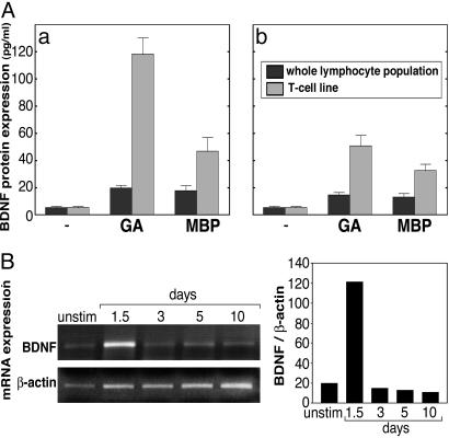
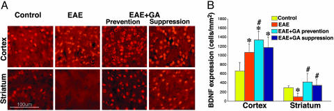
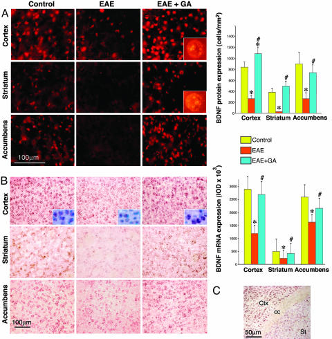
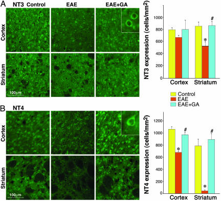
Neurotrophins (NTs) such as BDNF, NT-3, and NT-4 are important modulators of neuronal function and survival. Their expression in the CNS after various insults is thus of major therapeutic consequence. Glatiramer acetate [(GA) Copaxone], an approved drug for the treatment of multiple sclerosis, has been shown to induce Th2/3 cells that accumulate in the CNS, expressing in situ antiinflammatory cytokines and BDNF. In the present study, we investigated whether s.c. injections of GA, applied at various stages of experimental autoimmune encephalomyelitis, affect the expression of NTs, particularly BDNF, in the brain. In untreated experimental autoimmune encephalomyelitis mice, the expression of NTs was elevated shortly after disease appearance but subsequently declined below that of naive mice. In contrast, GA treatment led to sustained augmentation in the expression of BDNF, NT-3, and NT-4 in various brain regions as demonstrated by histological analysis of immunostained brain sections. GA treatment, even when started 45 days after disease induction, restored the impaired level of NTs to that of healthy mice. BDNF elevation after GA treatment was demonstrated on both protein and mRNA levels. Prominent staining was manifested not only by infiltrating GA-induced T cells, but also by CNS resident cells (neurons and astrocytes), indicative of a bystander therapeutic effect. Of importance, in GA-treated mice, intense BDNF expression was manifested by neuronal progenitors that migrated into lesions in injured regions. These results indicate that the immunomodulator GA exerts not only an antiinflammatory effect, but also enhances neuroprotection and regeneration of neural elements in the diseased brain.


Figures

Fig. 1.
Fig. 1.
Production of BDNF by GA-specific T cells. (A) BDNF protein secretion by whole lymphocyte populations and T cell lines isolated from brains (a) and spleens (b) of GA-immunized mice, in response to medium alone, GA (50 μg/ml), or MBP (100 μg/ml). BDNF concentrations were measured by ELISA. (B) RT-PCR analysis of BDNF and housekeeping β-actin mRNA from GA-specific T cell line, at various time points after exposure to GA.
Fig. 2.
Fig. 2.
BDNF expression. (Left) BDNF expression by immunostaining (red) in brains of EAE-induced mice at the peak of clinical symptoms, 22 days after induction, and the effect of GA treatment, starting either immediately after disease induction (prevention) or at the appearance of clinical manifestations at day 11 (suppression). (Right) Quantitative analysis of BDNF staining was performed by counting positively stained elements, three mice per treatment group, eight sections for each region. *, Significant effect over naive control; #, significant effect over EAE-untreated mice.
Fig. 3.
Fig. 3.
BDNF expression in brains of EAE-induced mice at the chronic disease phase, 60 days after induction, and the effect of GA treatment, starting at day 45 (delayed suppression). (A) Immunohistochemical analysis of BDNF protein level (red). (B) In situ hybridization of BDNF mRNA (pink) and nuclear staining (blue, Inset). Quantitative analysis of protein expression was performed by counting positively stained cells, and analysis of mRNA was performed by measuring integrated optical density. *, Significant effect over naive control; #, significant effect over EAE-untreated mice. (C) BDNF mRNA in coronal section depicting part of the cortex (Ctx), the striatum (st), and the corpus callosum (cc). Considerably less mRNA expression is observed in the striatum than in the cortex.
Fig. 4.
Fig. 4.
Immunohistochemical analysis of BDNF-expressing cells. (A) Characterization of BDNF+ cells (red) in GA-treated mice by double staining with specific subset markers (green): CD3, which depicts T cells shown in the talamus (a), glial fibrillary acidic protein (GFAP) for astrocytes in the corpus callosum (b), NeoN for neurons in the singulate cortex (c), and DCX for neuronal progenitors in the accumbens (d). (B) DCX+ neuronal progenitors (green) expressing BDNF (orange) in the SVZ of EAE (a) and EAE + GA (b) mice. (Insets) A larger proportion of cells in the GA-treated mice coexpressed BDNF. Neuroprogenitors expressing BDNF migrated from the SVZ toward lesions in the striatum (c) and the corpus callosum (d). Note the DCX-expressing fibers extending around the lesions. (C) Cytokine expression by BDNF-expressing cells; triple staining in the optic tract of a GA-treated mouse. Coexpression of BDNF (red), IL-10 (green), and lack of IFN-γ (blue). Arrows indicate double positive cells. Representative figures from four EAE and five EAE + GA mice are shown.
Fig. 5.
Fig. 5.
Expression of NTs by immunostaining (green) in the brains of EAE-induced mice at the chronic disease phase, 60 days after induction, and the effect of GA treatment, starting at day 45 (delayed suppression). NT3 (A) and NT4 (B) expression in the motor cortex (layers 2 and 3) and the striatum. Quantitative analysis was performed by counting positively stained cells, two to three mice per treatment group and eight sections for each region. *, Significant effect over naive control; #, significant effect over EAE-untreated mice.

References

    1. Behi, M. E., Dubucquoi, S., Lefranc, D., Zephir, H., De Seze, J., Vermersch, P. & Prin, L. (2005) Immunol. Lett. 96, 11-26. - PubMed
    1. Hellings, N., Raus, J. & Stinissen, P. (2002) Immunol. Res. 25, 27-51. - PubMed
    1. Bjartmar, C., Wujek, J. R. & Trapp, B. D. (2003) J. Neurol. Sci. 15, 165-171. - PubMed
    1. Lassmann, V., Gottmann, K. & Malcangio, M. (2003) Prog. Neurobiol. 69, 341-374. - PubMed
    1. Riley, C. P., Cope, T. C. & Buck, C. R. (2004) J. Mol. Histol. 35, 771-783. - PubMed

Publication types

MeSH terms