. 2005 Dec 27;102(52):19126-31.
doi: 10.1073/pnas.0508215102. Epub 2005 Dec 19.

Complex I deficiency primes Bax-dependent neuronal apoptosis through mitochondrial oxidative damage

Affiliations

Complex I deficiency primes Bax-dependent neuronal apoptosis through mitochondrial oxidative damage

Celine Perier et al. Proc Natl Acad Sci U S A. .

Abstract

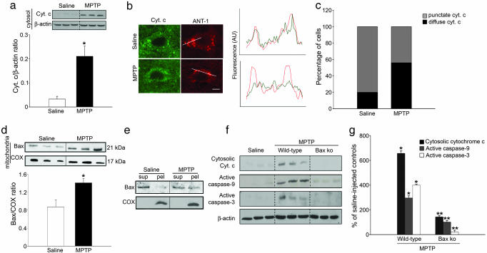
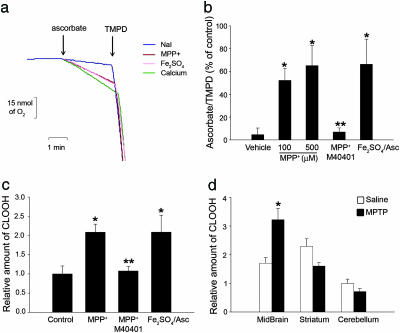
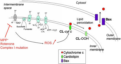
Dysfunction of mitochondrial complex I is a feature of human neurodegenerative diseases such as Leber hereditary optic neuropathy and Parkinson's disease. This mitochondrial defect is associated with a recruitment of the mitochondrial-dependent apoptotic pathway in vivo. However, in isolated brain mitochondria, complex I dysfunction caused by either pharmacological or genetic means fails to directly activate this cell death pathway. Instead, deficits of complex I stimulate intramitochondrial oxidative stress, which, in turn, increase the releasable soluble pool of cytochrome c within the mitochondrial intermembrane space. Upon mitochondrial permeabilization by the cell death agonist Bax, more cytochrome c is released to the cytosol from brain mitochondria with impaired complex I activity. Given these results, we propose a model in which defects of complex I lower the threshold for activation of mitochondrial-dependent apoptosis by Bax, thereby rendering compromised neurons more prone to degenerate. This molecular scenario may have far-reaching implications for the development of effective neuroprotective therapies for these incurable illnesses.


Figures

Fig. 1.
Fig. 1.
Bax-dependent recruitment of mitochondrial apoptotic pathway following complex I inhibition in mice. (a) Cytochrome c levels are increased in ventral midbrain cytosolic fractions of MPTP-intoxicated mice at day 4 after the last MPTP injection, as determined by immunoblot. (b and c) Double immunofluorescence of substantia nigra pars compacta (SNpc) sections with cytochrome c (green) and the mitochondrial marker ANT-1 (red) show that in saline injected animals, 80% of SNpc neurons (n = 77) exhibit cytochrome c immunostaining colocalized with ANT-1, indicative of its mitochondrial localization. After complex I inhibition by MPTP, ≈60% of SNpc neurons (n = 206) show a diffuse cytochrome c staining no longer colocalized with ANT-1, indicative of its cytosolic redistribution. The fluorescence intensity profiles reported in the diagram correspond to the lines drawn in the confocal images (see Materials and Methods for details) (AU, fluorescence arbitrary units). (d) Bax levels are increased in ventral midbrain mitochondrial fractions at day 4 after the last MPTP injection, indicating Bax mitochondrial translocation. (e) In saline-injected animals, most of mitochondrial Bax appears in the supernatant fraction (Sup) after alkaline extraction, indicating its loose association with mitochondrial membranes. In contrast, after complex I blockade by MPTP, a significant fraction of mitochondrial Bax remains in the mitochondrial pellet (Pel), indicating its insertion into mitochondrial membranes. The inner mitochondrial membrane protein cytochrome oxidase was not extracted by alkaline treatment. (f and g) Genetic ablation of Bax in mutant mice (Bax ko) attenuates MPTP-induced cytochrome c release and caspase activation. *, P < 0.05, compared with saline-injected mice; **, P < 0.05, compared with MPTP-injected wild-type mice.
Fig. 2.
Fig. 2.
Complex I inhibition stimulates ROS production and promotes Bax-dependent cytochrome c release in isolated brain mitochondria. (a) MPP+ induces a dose-dependent inhibition of complex I-driven mitochondrial respiration, as assessed by monitoring oxygen consumption after addition of ADP, which in normal mitochondria induces a transient mitochondrial depolarization with a subsequent burst of oxygen consumption (state 3 respiration) until the added ADP is converted to ATP (state 4). (b) Complex I inhibition by MPP+ induces dose-dependent ROS production in brain mitochondria, as assessed by measuring H2O2 using the fluorescent dye Amplex Red. (c) Complex I inhibition with 100 μM MPP+ or incubation with ≈100 nM recombinant Bax, alone did not trigger significant release of cytochrome c from isolated brain mitochondria. However, combining complex I inhibition with recombinant Bax resulted in a marked release (>60%) of cytochrome c. This effect was abolished by 50 μM of the superoxide dismutase mimetic M40401. Matrix mitochondrial protein HSP60 was not mobilized by any of the tested conditions. *, P < 0.05, compared with untreated mitochondria; **, P < 0.05, compared with mitochondria treated with MPP+ and recombinant Bax.
Fig. 3.
Fig. 3.
Genetic disruption of complex I in cybrid cells stimulates ROS production and promotes Bax-dependent cytochrome c release. (a) Isolated mitochondria from mutant 3460/ND1 cybrids (cybrid mt) exhibit reduced complex I-driven mitochondrial respiration, as assessed by monitoring oxygen consumption supported by NADH-linked substrates glutamate/malate. (b) Impairment of mitochondrial respiration in mutant 3460/ND1 cybrids is associated with an increased production of ROS that was quenched by 50 μM of M40401. (c) Recombinant Bax (≈100 nM) induced a marked release (≈50%) of cytochrome c in mitochondria isolated from mutant 3460/ND1 cybrids but not from wild-type cybrids (cybrid wt) or parental osteosarcoma cells. This effect was attenuated by 50 μM M40401. *, P < 0.05, compared with noncybrid and cybrid wild-type mitochondria; **, P < 0.05, compared with mutant cybrid mitochondria.
Fig. 4.
Fig. 4.
Complex I inhibition increases the soluble pool of cytochrome c in the mitochondrial intermembrane space by oxidizing cardiolipin. (a and b) Complex I inhibition by MPP+ induced a dose-dependent increase of mitochondrial ascorbate/TMPD-driven respiration ratio, consistent with an increased intermembrane soluble pool of cytochrome c. This effect was prevented by 50 μM M40401 and could be reproduced by the ROS-generating compound, Fe2SO4 (60 μM)/ascorbate (500 μM). Sodium iodide (NaI) was used as vehicle, because MPP+ was used in the form of MPP+-I. Ca2+-mediated mitochondrial swelling, which increases the soluble pool of cytochrome c (20), was used as a positive control. (c) Complex I inhibition by MPP+ induced oxidation of inner mitochondrial membrane cardiolipin in isolated brain mitochondria, as assessed by determining cardiolipin hydroperoxide (CLOOH) content by HPLC. Oxidation of cardiolipin was also produced by Fe2SO4/ascorbate and was attenuated by M40401. (d) Oxidized cardiolipin was detected in ventral midbrain samples from MPTP-intoxicated mice but not in regions devoided of MPTP-induced cell loss, such as striatum and cerebellum. *, P < 0.05, compared with controls; **, P < 0.05, compared with MPP+-treated mitochondria.
Fig. 5.
Fig. 5.
Proposed pathogenic scenario induced by complex I deficiency. Pharmacological or genetic inhibition of complex I disrupts mitochondrial respiration and stimulates the mitochondrial production of ROS. As a consequence, an array of molecules is likely oxidatively modified in response to complex I defect, including the inner mitochondrial membrane lipid cardiolipin. Cardiolipin peroxidation, in turn, affects the binding of cytochrome c to the mitochondrial inner membrane, leading to an increased soluble pool of cytochrome c in the intermembrane space. Consequently, upon permeabilization of the outer mitochondrial membrane by activated Bax, a larger amount of mitochondrial cytochrome c can be released, making it more likely for a compromised neuron to undergo apoptosis (see Discussion for more details).

References

    1. Schon, E. A. & Manfredi, G. (2003) J. Clin. Invest. 111, 303-312. - PMC - PubMed
    1. Wallace, D. C., Singh, G., Lott, M. T., Hodge, J. A., Schurr, T. G., Lezza, A. M. S., Elsas, L. J. & Nikoskelainen, E. K. (1988) Science 242, 1427-1430. - PubMed
    1. Parker, W. D., Jr., Boyson, S. J. & Parks, J. K. (1989) Ann. Neurol. 26, 719-723. - PubMed
    1. Schapira, A. H., Cooper, J. M., Dexter, D., Clark, J. B., Jenner, P. & Marsden, C. D. (1990) J. Neurochem. 54, 823-827. - PubMed
    1. Dauer, W. & Przedborski, S. (2003) Neuron 39, 889-909. - PubMed

Publication types

MeSH terms