. 2005 Dec 27;102(52):19198-203.
doi: 10.1073/pnas.0505203102. Epub 2005 Dec 19.

Brain estrogen deficiency accelerates Abeta plaque formation in an Alzheimer's disease animal model

Affiliations

Brain estrogen deficiency accelerates Abeta plaque formation in an Alzheimer's disease animal model

Xu Yue et al. Proc Natl Acad Sci U S A. .

Abstract

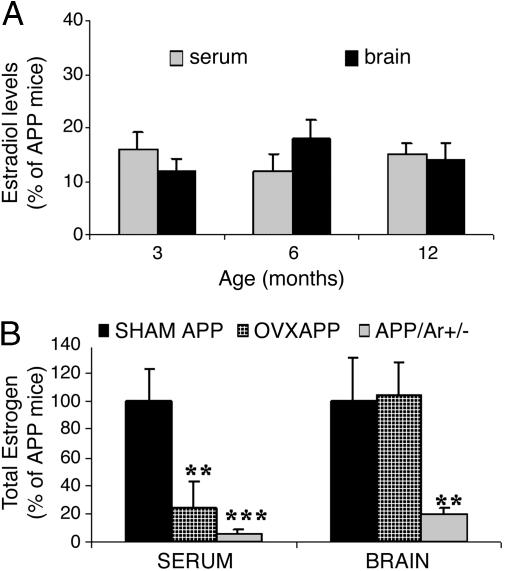
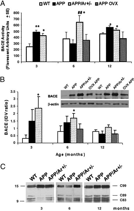
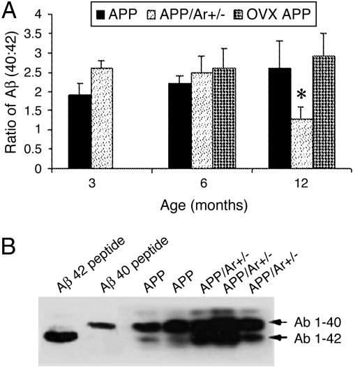
Much evidence indicates that women have a higher risk of developing Alzheimer's disease (AD) than do men. The reason for this gender difference is unclear. We hypothesize that estrogen deficiency in the brains of women with AD may be a key risk factor. In rapidly acquired postmortem brains from women with AD, we found greatly reduced estrogen levels compared with those from age- and gender-matched normal control subjects; AD and control subjects had comparably low levels of serum estrogen. We examined the onset and severity of AD pathology associated with estrogen depletion by using a gene-based approach, by crossing the estrogen-synthesizing enzyme aromatase gene knockout mice with APP23 transgenic mice, a mouse model of AD, to produce estrogen-deficient APP23 mice. Compared with APP23 transgenic control mice, estrogen-deficient APP23 mice exhibited greatly reduced brain estrogen and early-onset and increased beta amyloid peptide (Abeta) deposition. These mice also exhibited increased Abeta production, and microglia cultures prepared from the brains of these mice were impaired in Abeta clearance/degradation. In contrast, ovariectomized APP23 mice exhibited plaque pathology similar to that observed in the APP23 transgenic control mice. Our results indicate that estrogen depletion in the brain may be a significant risk factor for developing AD neuropathology.


Figures

Fig. 1.
Fig. 1.
Estrogen levels in mice. (A) Total 17β-estradiol was detected by RIA in the serum and brain from APP23/Ar+/- (n = 10) and APP23 (n = 10) mice at various ages. (B) Twelve APP23 mice were OVX or sham-operated at 3 months of age. By 6 months of age, levels of total 17β-estradiol were measured, in both serum and brain from APP23/Ar+/- (n = 10), OVX APP (n = 7), SHAM APP23 (n = 5), or naïve APP23 (n = 10) mice. *, P < 0.01; **, P < 0.001 (compared with naïve APP23 control mice).
Fig. 2.
Fig. 2.
The plaque formations in the brains of 6- and 12-month-old mice. Fourteen APP23 mice were OVX at 3 months of age. At age of 6 or 12 months, animals were killed and brains were prepared for immunohistochemistry. Shown is anti-Aβ (1-17) (6E10 clone antibody) immunostaining of sagittal brain sections from APP23 (n = 10; A and D), OVX APP23 (n = 7; B and E), and APP23/Ar+/- (n = 10; C and F) mice aged 6 (A-C) and 12 (D-F) months.
Fig. 3.
Fig. 3.
Expression of BACE activity and protein in mice with age. At 3, 6, or 12 months of age, brain from APP23 (n = 10) and APP23/Ar+/- (n = 13) mice were prepared for enzyme activity and Western blot. The OVX APP mice (n = 14) were OVX at 3 months, and brains were harvested only at 6 or 12 months of age. (A) BACE activity was detected by detection of fluorescent-labeled peptides. (B) BACE protein levels were measured by Western blot by using monoclonal anti-BACE and were normalized according to those of a β-actin control. Bars indicate the mean ratio of BACE/β-actin integrated optical density values (IDV). (Inset) Western blot of 6-month-old mice. (C)Representative immunoblots were immunostained with APPC8, an antibody against the APP C-terminal fragment C99 that results from BACE cleavage of APP, in WT, APP23, and APP23/Ar+/- mice at various ages. *, P < 0.05; **, P < 0.01 (compared with WT mice). ##, P < 0.05 (compared with APP23 mice).
Fig. 4.
Fig. 4.
Effect of genotypes on Aβ levels in the brain. (A) Ratio of Aβ40:Aβ42. Formic acid-extractable Aβ was measured in the mice. ELISA was used for detection of Aβ40:Aβ42 ratio in APP23 and APP23/Ar+/- mice. At various ages, brain tissues were harvested from APP23 (n = 10), APP23/Ar+/- (n = 13), and OVX APP (n = 7) mice and homogenized and prepared for ELISA as suggested by the manufacturer. Bars indicate the mean value for the experimental group. All values represent the average of four sets of duplicate determinations. *, P < 0.05 compared with APP23 mice. (B) Urea gel/Western blot analysis of the levels of Aβ40 and Aβ42 in APP23 (n = 8) and APP23/Ar+/- (n = 8) mice at 12 months of age. Synthetic peptide Aβ40 and Aβ42 were used as positive controls.

References

    1. Evans, D. A., Funkenstein, H. H., Albert, M. S., Scherr, P. A., Cook, N. R., Chown, M. J., Hebert, L. E., Hennekens, C. H. & Taylor, J. O. (1989) J. Am. Med. Assoc. 262, 2551-2556. - PubMed
    1. Haass, C. & De Strooper, B. (1999) Science 286, 916-919. - PubMed
    1. Vassar, R., Bennett, B. D., Babu-Khan, S., Kahn, S., Mendiaz, E. A., Denis, P., Teplow, D. B., Ross, S., Amarante, P., Loeloff, R., et al. (1999) Science 286, 735-741. - PubMed
    1. Dewachter, I., van Dorpe, J., Spittaels, K., Tesseur, I., Van Den Haute, C., Moechars, D. & Van Leuven, F. (2000) Exp. Gerontol. 35, 831-841. - PubMed
    1. Holsinger, R. M., McLean, C. A., Beyreuther, K., Masters, C. L. & Evin, G. (2002) Ann. Neurol. 51, 783-786. - PubMed

Publication types

MeSH terms

LinkOut - more resources