. 2005 Dec 27;102(52):18773-84.
doi: 10.1073/pnas.0509487102. Epub 2005 Dec 19.

On the mechanism of sensing unfolded protein in the endoplasmic reticulum

Affiliations

On the mechanism of sensing unfolded protein in the endoplasmic reticulum

Joel J Credle et al. Proc Natl Acad Sci U S A. .

Abstract

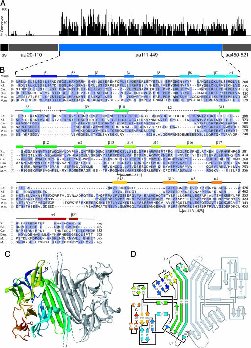
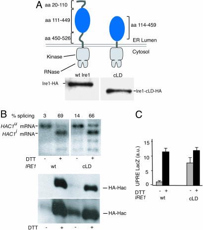
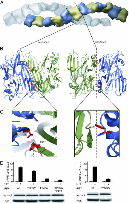
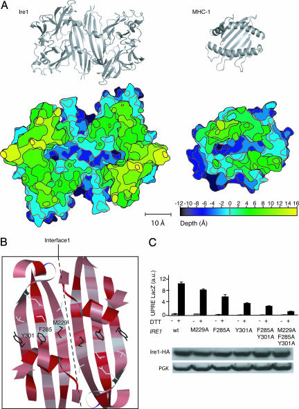
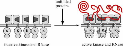
Unfolded proteins in the endoplasmic reticulum (ER) activate the ER transmembrane sensor Ire1 to trigger the unfolded protein response (UPR), a homeostatic signaling pathway that adjusts ER protein folding capacity according to need. Ire1 is a bifunctional enzyme, containing cytoplasmic kinase and RNase domains whose roles in signal transduction downstream of Ire1 are understood in some detail. By contrast, the question of how its ER-luminal domain (LD) senses unfolded proteins has remained an enigma. The 3.0-A crystal structure and consequent structure-guided functional analyses of the conserved core region of the LD (cLD) leads us to a proposal for the mechanism of response. cLD exhibits a unique protein fold and is sufficient to control Ire1 activation by unfolded proteins. Dimerization of cLD monomers across a large interface creates a shared central groove formed by alpha-helices that are situated on a beta-sheet floor. This groove is reminiscent of the peptide binding domains of major histocompatibility complexes (MHCs) in its gross architecture. Conserved amino acid side chains in Ire1 that face into the groove are shown to be important for UPR activation in that their mutation reduces the response. Mutational analyses suggest that further interaction between cLD dimers is required to form higher-order oligomers necessary for UPR activation. We propose that cLD directly binds unfolded proteins, which changes the quaternary association of the monomers in the membrane plane. The changes in the ER lumen in turn position Ire1 kinase domains in the cytoplasm optimally for autophosphorylation to initiate the UPR.


Figures

Fig. 1.
Fig. 1.
The Ire1 cLD. (A) The relative conservation of amino acids is plotted along the sequence of Ire1 LD. The blue bar represents the cLD, the structure of which is shown below. The gray bars represent regions that were disordered in LD crystals and absent in cLD crystals. The black bar represents the signal sequence (ss). (B) Amino acid alignment of IRE1 and PERK LDs. (S.c., Saccharomyces cerevisiae; K.l., Kluveromyces lactis; C.e., Caenorhabditis elegans; D.m., Drosophila melanogaster; M.m., Muscus musculus-a; I, Ire1 cLD; P, PERK cLD. Conservation of residues among species was scored by using blossum62 (46). Blue represents residues of high conservation. Secondary structural elements are indicated above the alignment and correspond in color to those of the ribbon diagram of the Ire1 cLD in C. Dashed lines (L1 and L2) represent regions found disordered in the structure. The asterisks mark residues that have been mutated in this study. For each sequence, amino acid number 1 is the initiating Met. The D.m. sequence is incorrect in the databases; an in-house resequenced sequence is used in the alignment (Julie Hollien and Jonathan Weissman, personal communication). The PERK sequence has two additional insertions (amino acids 286–314 and 413–428) where indicated. (C) Ribbon diagram of the cLD dimer as seen in the asymmetric unit corresponding to residues 111–449 have been colored with a rainbow gradient with from N terminus (blue) to C terminus (red). (D) Schematic connectivity diagram (road map) of the cLD using the same coloring scheme as in B and C.
Fig. 2.
Fig. 2.
Functional analysis of cLD-Ire1. (A) (Upper) Topography of Ire1 and cLD-Ire1. The Ire1 cLD construct contains an ER-LD starting with amino acid 114 and ending in amino acid 449. (Lower) Immunoblot of HA-tagged Ire1 and cLD-Ire1. (B) Northern blot analysis of HAC1 mRNA in control and DTT-treated cells expressing wild-type Ire1 or cLD-Ire1. Unspliced HAC1u and spliced HAC1i mRNAs are indicated; the lower bands are splicing intermediates. Immunoblot against Hac1-HA in control or DTT-treated cells expressing wild-type (wt) Ire1 or cLD-Ire1. A short exposure (Upper) and a long exposure (Lower) are shown. (C) LacZ activity assay in control cells (gray bars) and DTT-treated cells (black bars) expressing wild-type Ire1 or cLD-Ire1.
Fig. 3.
Fig. 3.
Analysis of Interfaces 1 and 2. (A) Surface representation the unit cell of cLD monomers in space group P6522. There are 24 cLD monomers per unit cell, arranged in two strands that twist around the 65 axis. Dashed lines represent the position of interfaces 1 and 2 within the strand between monomers. (B) Ribbon diagram of cLD dimers connected through Interface 1 (Left) or Interface 2 (Right). Dashed lines represent the interfaces between the twofold symmetrical dimers as seen in the asymmetric unit. The red residues shown in stick representation have been mutated. (C) Enlarged view of residues that were mutated (T226W and F247A in Interface 1 and W426A in Interface 2). (D) LacZ activity assay in control cells (gray bars) and DTT-treated cells (black bars) expressing wild-type Ire1 and Ire1 with the indicated mutations. Lower shows an immunoblot of Ire1-HA and its mutant forms.
Fig. 4.
Fig. 4.
Analysis of the central groove in cLD dimers. (A) (Upper) Ribbon diagrams of the cLD dimer (Left) and MHC-1 (Right) shown in the same scale for comparison. Note that the slant of the β-strands is opposite between cLD and MHC. (Lower) A topographic map of cLD and MHC-1 seen from the top. The map displays the grooves as deep canyons of roughly equivalent depths and widths in the two structures. The vertical spacing of the contour lines connecting points of equal depths is 2 Å, and different elevations are colored according to the scale provided. The red index line at depth = 0 is set in both structures at the point were the rim becomes discontinuous. Relative to this contour, the grooves in both structures are 11-Å deep at their lowest point. (B) Ribbon representation looking into the cLD groove, displaying the residues mutated. The ribbon drawing is colored by amino acid conservation. Red corresponds to phylogenetically conserved amino acids. Note the “candy cane” pattern of conserved residues pointing into the groove. (C)(Upper) LacZ activity assay in control cells (gray bars) and DTT-treated cells (black bars) expressing wild-type Ire1 or Ire1 with the indicated mutations. (Lower) Immunoblot of Ire1-HA and its mutant forms.
Fig. 5.
Fig. 5.
Model for unfolded protein recognition by Ire1. The model depicts Ire1 activation through oligomerization brought about by binding of unfolded proteins (indicated in red). Direct or indirect interactions between unfolded protein chains may contribute to activation. On the ER-luminal side of the membrane, the postulated unfolded protein-binding groove formed by Ire1 cLD dimerization through Interface 1 is indicated in dark gray. On the cytoplasmic side of the ER membrane, oligomerization juxtaposes the Ire1 kinase domains, which undergo a conformational change after autophosphorylation that activates the RNase function of Ire1. Inactive Ire1 could either be monomeric as shown or exist already in oligomeric yet inactive states whose quaternary associations change upon unfolded protein binding.

Similar articles

Cited by

References

    1. Gething, M.-J. & Sambrook, J. (1992) Nature 355, 33–45. - PubMed
    1. Stevens, F. J. & Argon, Y. (1999) Semin. Cell Dev. Biol. 10, 443–454. - PubMed
    1. Cox, J. S., Shamu, C. E. & Walter, P. (1993) Cell 73, 1197–1206. - PubMed
    1. Patil, C. & Walter, P. (2001) Curr. Opin. Cell Biol. 13, 349–355. - PubMed
    1. Ron, D. (2002) J. Clin. Invest. 110, 1383–1388. - PMC - PubMed

Publication types

MeSH terms

Substances

Associated data