. 2006 Feb;16(2):240-50.
doi: 10.1101/gr.4571606. Epub 2005 Dec 19.

L1 integration in a transgenic mouse model

Affiliations

L1 integration in a transgenic mouse model

Daria V Babushok et al. Genome Res. 2006 Feb.

Abstract

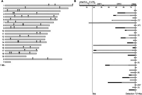
To study integration of the human LINE-1 retrotransposon (L1) in vivo, we developed a transgenic mouse model of L1 retrotransposition that displays de novo somatic L1 insertions at a high frequency, occasionally several insertions per mouse. We mapped 3' integration sites of 51 insertions by Thermal Asymmetric Interlaced PCR (TAIL-PCR). Analysis of integration locations revealed a broad genomic distribution with a modest preference for intergenic regions. We characterized the complete structures of 33 de novo retrotransposition events. Our results highlight the large number of highly truncated L1s, as over 52% (27/51) of total integrants were <1/3 the length of a full-length element. New integrants carry all structural characteristics typical of genomic L1s, including a number with inversions, deletions, and 5'-end microhomologies to the target DNA sequence. Notably, at least 13% (7/51) of all insertions contain a short stretch of extra nucleotides at their 5' end, which we postulate result from template-jumping by the L1-encoded reverse transcriptase. We propose a unified model of L1 integration that explains all of the characteristic features of L1 retrotransposition, such as 5' truncations, inversions, extra nucleotide additions, and 5' boundary and inversion point microhomologies.


Figures

Figure 1.
Figure 1.
Structures of L1 element and L1-mless transgene. (A) Human full-length L1 contains a 5′UTR, ORF1, ORF2, 3′UTR, and a poly(A) tail. (B) L1LRE3 is tagged in its 3′UTR with a markerless retrotransposition cassette (mless) and is driven by the mouse Hsp70-2 promoter. Mless contains the human γ-globin intron in the same orientation as L1 (SD and SA denote splice donor and acceptor sites, respectively), flanked by splice acceptors from the human Bcl-2 gene (not shown). After retrotransposition, the intron is spliced, allowing de novo insertions to be distinguished from the original transgene by PCR with oligos L16045(30) and mlessfor2 (black arrows). The expected band pattern from this “genotyping PCR” is shown on the right. Gray arrows SP1–SP4 indicate insertion-specific oligos used in TAIL–PCR.
Figure 2.
Figure 2.
Characterization of a pedigree segregating transgene-11. (A)F1 mouse and its 15 offspring (top pedigree schematic) were genotyped by PCR with L16045(30) and mlessfor2 oligos (top gel) to reveal 11/15 F2s carrying transgene (1134 bp) and 7/15 carrying de novo inserts (231 bp). Minus DNA control was negative; de novo insertion presence was confirmed with different oligos and independently isolated tail DNA stocks (data not shown). All transgene inheritance was accounted for by transgene-11-specific PCR (middle gel, 446 bp). Southern blot (bottom gel) of 10 μg of tail DNA, digested with AvrII, and probed with a 32P-labeled 376-bp probe showed multicopy (3850 bp) and single-copy (2764 bp) fragments expected for AvrII digest of transgene-11, shown in B. No other fragments were detected, suggesting that the frequency of de novo insertions is less than one copy/cell. Four F2s contained inserts characterized by TAIL–PCR (gray shading in pedigree schematic). A 1-kb Plus DNA Ladder is shown in lane 1 of all gels. (B) A hypothetical multicopy transgene-11 array with relevant AvrII sites and the 376 bp probe (black rectangle).
Figure 3.
Figure 3.
Genomic distribution and structural features of de novo insertions. (A) Using TAIL–PCR, 51 de novo integration sites were determined, 48 of which were uniquely mapped to the mm6 assembly of the mouse genome. Depicted are the locations of 47 inserts (one mapped to an unplaced location and is not shown). 1-Mb scale reference is shown at the bottom. (B) The structures of 33 fully characterized de novo inserts. A hypothetical full-length (FL) insert is shown at the top, with vertical lines indicating the 6-kb endogenous FL element and the 214-bp detection limit in our study. De novo integrants are shown on separate lines, aligned to the FL element. (Direct fragment) Gray rightward arrow; (inverted fragment) black rectangle; (deletion of sequence in inverted elements) white rectangle; (extra 5′ nt) numbers in parentheses; (dual inversions) asterisk. Three elements mobilized ∼6 kb of sequence, one is FL and two are nearly FL.
Figure 4.
Figure 4.
Characteristic features of L1 retrotransposition in de novo integrants. (A) Average frequencies of each base in the 10 nt surrounding the insertion sites of de novo inserts. (B) Insertion length distribution of 33 fully characterized integrants. Lengths of the inverted inserts were calculated by adding the lengths of inverted and direct segments. (C) The distribution of TSD lengths among 33 fully characterized inserts. (D) Observed microhomology (MH) frequencies at 5′ boundaries of noninverted 5′-truncated insertions (black), and at the inversion points (white) and 5′ boundaries (gray) of insertions containing inversions. Observed 5′MH frequencies were in agreement with those in cultured cells (Symer et al. 2002) (diagonal stripes), and were higher than expected for random ligation (Roth et al. 1985) using both the random nucleotide distribution (data not shown) and the nucleotide distribution in the 30-bp downstream of the insertion site (horizontal stripes).
Figure 5.
Figure 5.
(A) 5′ junctions of insertions containing 26 and 47 extra 5′ nucleotides. Their likely formation is depicted in B and C, respectively. TSD bases are in lower case. (Flanking host DNA) Solid black lines; (L1 RNA) gray line; (nascent cDNA strand) light-green line; (conceptual base pairing) dotted black lines. (B) A schematic of the “dual inversion” mechanism that created the 26 extra nucleotides between the flanking host DNA and the 238-bp 5′-truncated insert. Extra nucleotides are a product of two successive twin priming reactions (in A, highlighted in light and dark blue). Likely, the first twin priming reaction was initiated using a 1-bp MH, synthesizing the first inverted fragment of 18 bp (light blue). Then, the RT underwent a template jump, creating the second inverted fragment of 8 bp (dark blue). (C) Successive L1 RT template jumps created an insertion of 47 nt in a 368-bp de novo integrant. The 47-nt addition contains three overlapping regions of homology to the host DNA immediately 5′ to the insertion site (in A, underlined orange, bold and yellow, and italicized and blue). When L1 RT reached the end of the RNA template at the end of a TPRT reaction, it likely template jumped from L1 RNA to the host DNA strand. Nascent cDNA was elongated by 11 bases (orange) copying a portion of host DNA. A second RT jump occurred, copying the same region of host DNA for 26 nt (yellow). One base was added (data not shown), and a third RT template jump occurred extending for seven more nucleotides (blue). Two bases were added (data not shown), and the structure was resolved, creating an addition of 47 nt. (D) Model for L1 integration. (Flanking host DNA) Solid black lines; (L1 RNA) gray line; (nascent cDNA strand) green line; (nascent 2nd strand) blue line; (homologous recombinational repair) orange crossed lines; (recombination products [no sequence changes expected]) stippled black/yellow and black/blue lines; (5′MH-guided base pairing) dotted red line; (TSD) target site duplication. See Discussion for detailed explanation.

References

    1. Bibillo, A. and Eickbush, T.H. 2002a. High processivity of the reverse transcriptase from a non-long terminal repeat retrotransposon. J. Biol. Chem. 277 34836-34845. - PubMed
    1. ———. 2002b. The reverse transcriptase of the R2 non-LTR retrotransposon: Continuous synthesis of cDNA on non-continuous RNA templates. J. Mol. Biol. 316 459-473. - PubMed
    1. ———. 2004. End-to-end template jumping by the reverse transcriptase encoded by the R2 retrotransposon. J. Biol. Chem. 279 14945-14953. - PubMed
    1. Boissinot, S., Chevret, P., and Furano, A.V. 2000. L1 (LINE-1) retrotransposon evolution and amplification in recent human history. Mol. Biol. Evol. 17 915-928. - PubMed
    1. Boissinot, S., Entezam, A., and Furano, A.V. 2001. Selection against deleterious LINE-1-containing loci in the human lineage. Mol. Biol. Evol. 18 926-935. - PubMed

Publication types

Substances

LinkOut - more resources