. 2006 Jan 1;176(1):472-83.
doi: 10.4049/jimmunol.176.1.472.

Helminth-primed dendritic cells alter the host response to enteric bacterial infection

Affiliations

Helminth-primed dendritic cells alter the host response to enteric bacterial infection

Chien-Chang Chen et al. J Immunol. .

Abstract

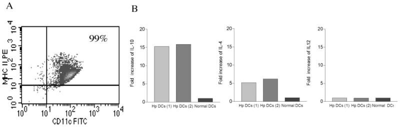
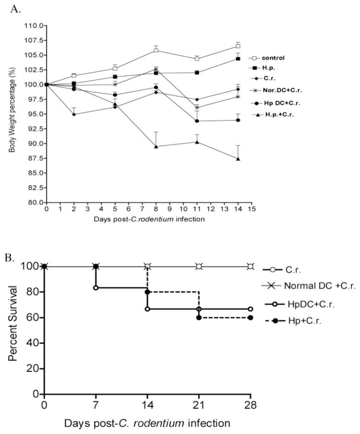
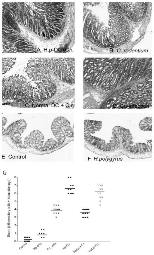
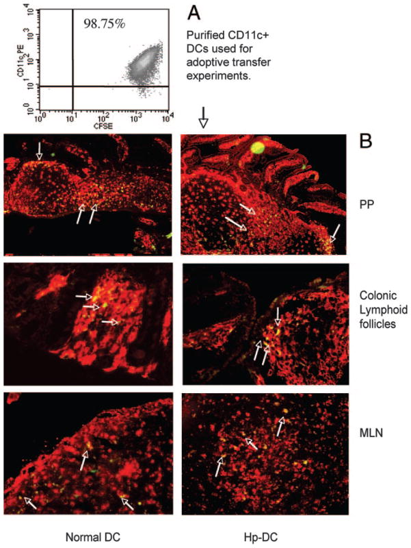
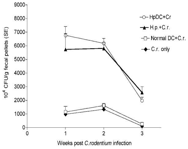
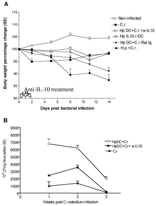
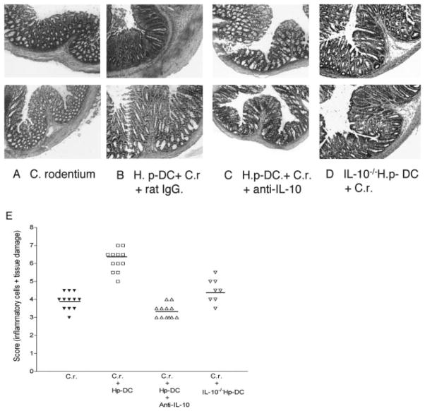
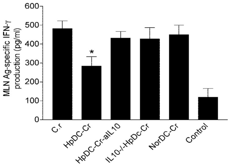
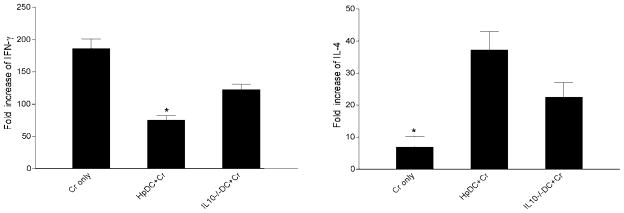
To examine whether intestinal helminth infection may be a risk factor for enteric bacterial infection, a murine model was established using the intestinal helminth Heligomosomoides polygyrus and a murine pathogen Citrobacter rodentium, which causes infectious colitis. Using this model we recently have shown that coinfection with the Th2-inducing H. polygyrus and C. rodentium promotes bacterial-associated disease and colitis. In this study, we expand our previous observations and examine the hypothesis that dendritic cells (DC) stimulated by helminth infection may play an important role in the regulation of the intestinal immune response to concurrent C. rodentium infection as well as in the modulation of the bacterial pathogenesis. We show that H. polygyrus infection induces DC activation and IL-10 expression, and that adoptive transfer of parasite-primed DC significantly impairs host protection to C. rodentium infection, resulting in an enhanced bacterial infection and in the development of a more severe colonic injury. Furthermore, we demonstrate that adoptive transfer of parasite-primed IL-10-deficient DCs fails to result in the development of a significantly enhanced C. rodentium-mediated colitis. Similarly, when the DC IL-10 response was neutralized by anti-IL-10 mAb treatment in mice that received parasite-primed DC, no deleterious effect of the parasite-primed DC on the host intestinal response to C. rodentium was detected. Thus, our results provide evidence to indicate that the H. polygyrus-dependent modulation of the host response to concurrent C. rodentium infection involves IL-10-producing DCs.


Conflict of interest statement

Disclosures

The authors have no financial conflict of interest.

Figures

FIGURE 1
FIGURE 1
More severe disease pathology is shown in coinfected mice. Coinfection with H. polygyrus and C. rodentium induces significant body weight loss (A) and colonic inflammation (B). BALB/c mice were infected with H. polygyrus (200 L3) and inoculated with C. rodentium (5 × 10⁸ CFU) 7 days later. A, Body weight changes of mice that were infected with C. rodentium (○), H. polygyrus (■), both H. polygyrus and C. rodentium (▲), and normal control (□) mice during the course of the experiment (2 wk) are shown. Data shown are pooled from three independent experiments and are expressed as the body weight change as a percentage of the individual mouse initial body weight ± SE (n = 10–15) at each time point. B, Coinfection with intestinal helminth parasite promotes C. rodentium-mediated colonic injury. Colon tissue was removed from uninfected mice or mice infected with H. polygyrus, C. rodentium, or both at 2 wk postbacterial infection, frozen in Tissue-Tek OCT compound and stained with H&E. Magnification, ×100. C and D, Helminth coinfection promotes C. rodentium-associated CD4+ T cell infiltration into colonic LP. E and F, Helminth coinfection results in an expansion of CD11c+ DCs in colonic lymphoid follicle. Five-micrometer sections of frozen colonic tissue (in OCT) were cut and fixed in ice-cold acetone. After washing with PBS, the sections were blocked with PBS-1% BSA. The tissue sections were incubated with anti-CD4 FITC (green) and anti-CD11c Ab, followed by Cy3-labeled hamster IgG. The sections were analyz+ ed by immunofluorescent microscopy. All images were digitized and cropped in Adobe Photoshop LE 5.0 (Adobe Systems). C and E, The mean number of positive cells is detected in each high-power field (×200) by counting 10 fields from each sample (samples from three mice per group were counted).
FIGURE 2
FIGURE 2
H. polygyrus induces DC IL-10 expression. A, FACS data show the purified CD11c+ DCs are MHC class II-positive. B, Cytokine expression of CD11c+ DCs (pooled from spleen and MLN) from H. polygyrus-infected mice by real-time PCR. Two preparations of DCs obtained from H. polygyrus-infected mice are shown. Values are mean fold increase compared with the baseline obtained from normal control animals.
FIGURE 3
FIGURE 3
Adoptive transfer of H. polygyrus-primed CD11c+ DCs induces body weight loss and enhances C. rodentium-induced disease status. A, Body weight changes of mice that were infected with C. rodentium (◆), with H. polygyrus (■), H. polygyrus-primed DC recipient mice infected with C. rodentium (●), normal DC recipient mice infected with C. rodentium (*), normal DC recipient mice infected with both H. polygyrus and C. rodentium (▲), and normal control (□) during the course of the experiments. Data shown are pooled from three experiments and are the percentage of initial body weight ± SE (n = 10–15) at each time point). B, Survival curve. There was no mortality in C. rodentium-infected mice or mice that received normal DC. However, there is a 20% mortality at 2 wk postbacterial infection in mice that received parasite-primed DCs.
FIGURE 4
FIGURE 4
Colonic histology of different groups at 2 wk postbacterial infection. A, Mice received CD11c+ DCs isolated from H. polygyrus-infected mice were then infected with C. rodentium. B, C. rodentium-infected mice. C, Mice adoptively transferred with CD11c+ DCs from normal BALB/c mice were then infected with C. rodentium. D, Mice coinfected with H. polygyrus and C. rodentium. E, Normal mice. F, H. polygyrus-infected mice. The results show that adoptive transfer of parasite-primed CD11c+ DCs enhances C. rodentium-mediated colonic pathology. G, Disease score of colonic inflammation is shown. The scores are assessed by determination of infiltration of inflammatory cells (range from 0 to 4), together with the evaluation of colon tissue damage (also a range from 0 to 4), with 0 scored normal and a score of 4 showing the most disease. The data shown are measurements of individual mouse (n = 8–12 per group) pooled from two to three independent experiments.
FIGURE 5
FIGURE 5
Detection of the adoptively transferred DCs in GALT of recipient mice. A, FACS data show CFSE-labeled CD11c+ DCs used for the adoptive transfer experiments. B, Detection of CFSE-labeled (green) CD11c+ DCs (Cy3, red) in the PP, MLN, and colonic lymphoid follicle. Arrows indicate some of the double stained cells (yellow). CD11c+ DCs were purified from H. polygyrus-infected (7 days postinfection), and normal mice and were adoptively transferred to normal BALB/c mice. The recipient mice were sacrificed 3 days later. The tissue sections (PP, MLN, colon) were prepared and stained with anti-CD11c-biotin and followed by streptavidin-Cy3. The sections were analyzed by immunofluorescent microscopy.
FIGURE 6
FIGURE 6
Mice that received H. polygyrus-primed DCs and then infected with C. rodentium have a higher bacterial output in the fecal pellets. The data shown are the number of bacteria recovered from fecal samples of C. rodentium-infected mice and mice with adoptive transfer of DCs at 1, 2, and 3 wk postinfection. The data are represented as the mean ± SEM (n = 5–10 mice) at each time point.
FIGURE 7
FIGURE 7
Alterations induced by adoptive transfer of parasite-primed DCs in mice can be reversed by anti-IL-10 treatment at the time of C. rodentium infection. A, Body weight changes of mice that were infected with C. rodentium (■), received H. polygyrus-primed DC and were then infected with C. rodentium treated with control Ab (▼), parasite-primed DC recipients, infected with C. rodentium and treated with anti-IL-10 (□), infected with both H. polygyrus and C. rodentium (●), adoptively transferred DCs obtained from IL-10-deficient, H. polygyrus-infected mice (△) and normal control (○) during the course of the experiments. Data shown are pooled from two to three experiments and are the percentage of change of initial body weight ± SE (n = 10–15 mice) at each time point. B, C. rodentium output in fecal pellets. The data shown are the number of bacteria recovered from fecal samples of C. rodentium-infected mice (◆) with adoptive transfer of DCs (○) and treated with anti IL-10 (●) at 1, 2, and 3 wk postinfection. The data are represented as the mean ± SEM (n = 5–10 mice) at each time point.
FIGURE 8
FIGURE 8
Colonic histopathology shows that adoptive transfer of CD11c+ DCs that lack IL-10 fails to promote C. rodentium-induced colitis. A, Colon section from C. rodentium-infected mice. B, Colon section of parasite-primed DC recipient with C. rodentium infection. C, Colonic histopathology of mice that received parasite-primed DC infected with C. rodentium and treated with anti-IL-10 mAb. D, Colon section of the parasite-primed IL-10-deficient (IL-10/) DC recipient infected with C. rodentium. Duplicate samples are presented from each group. E, Disease score of colonic inflammation as described in Fig. 3.
FIGURE 9
FIGURE 9
Adoptive transfer of helminth-primed DC results in an inhibition of bacterial Ag-specific IFN-γ production by MLN. MLN cells were cultured with and without C. rodentium Ag (10 μg/ml) for 72 h. Th1 (IFN-γ and Th2 (IL-4) cytokine production was measured using ELISA. The data show that C. rodentium infection induces Ag-specific IFN-γ production and that adoptive transfer of DCs from helminth-infected mice causes a significant suppression of bacteria-specific IFN-γ response. No detectable bacteria-specific Th2 (IL-4) response was found in any groups (data not shown).
FIGURE 10
FIGURE 10
Cytokine mRNA expression (IFN-γ and IL-4) in colon tissue as measured by real-time PCR at 2 wk postbacterial infection. Values are mean fold increase compared with the baseline obtained from normal control animals. The data shown are from one of two experiments performed, showing similar results. *, p < 0.05 (n = 3–5 mice per group).

Similar articles

Cited by

References

    1. Nokes C, Cooper ES, Robinson BA, Da B. Geohelminth infection and academic assessment in Jamaican children. Trans R Soc Trop Med Hyg. 1991;85:272–273. - PubMed
    1. Crompton DWT, Nesheim MC. Nutritional impact of intestinal helminthiasis during the human life cycle. Annu Rev Nutr. 2002;22:35–59. - PubMed
    1. Yazdanbakhsh M, Kremsner PG, van Ree R. Allergy, parasites, and the hygiene hypothesis. Science. 2002;296:490–494. - PubMed
    1. Elliott DE, Li J, Blum A, Metwali A, Qadir K, Urban JF, Jr, Weinstock JV. Exposure to schistosome eggs protects mice from TNBS-induced colitis. Am J Physiol. 2003;284:G385–G391. - PubMed
    1. Fox JG, Beck P, Dangler CA, Whary MT, Wang TC, Shi HN, Nagler-Anderson C. Concurrent enteric helminth infection modulates inflammation and gastric immune responses and reduces helicobacter-induced gastric atrophy. Nat Med. 2000;6:536–542. - PubMed

Publication types

MeSH terms