

Direct pyrogenic input from prostaglandin EP3 receptor-expressing preoptic neurons to the dorsomedial hypothalamus
- PMID: 16367780
- PMCID: PMC2441892
- DOI: 10.1111/j.1460-9568.2005.04515.x
Direct pyrogenic input from prostaglandin EP3 receptor-expressing preoptic neurons to the dorsomedial hypothalamus
Abstract
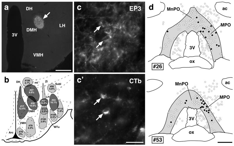
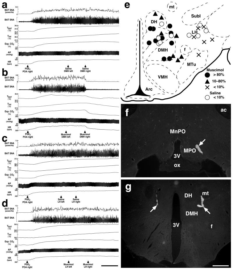
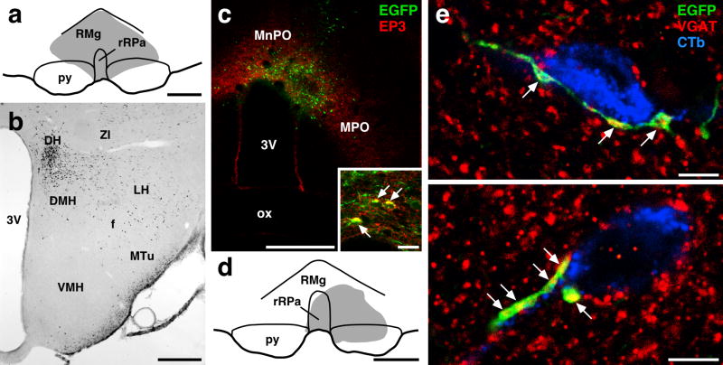
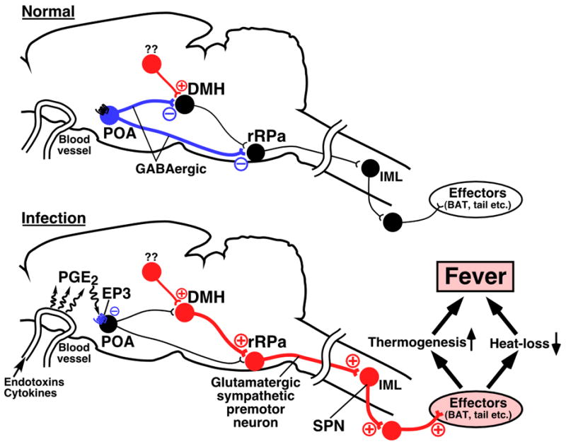
Fever is induced by a neuronal mechanism in the brain. Prostaglandin (PG) E2 acts as a pyrogenic mediator in the preoptic area (POA) probably through the EP3 subtype of PGE receptor expressed on GABAergic neurons, and this PGE2 action triggers neuronal pathways for sympathetic thermogenesis in peripheral effector organs including brown adipose tissue (BAT). To explore pyrogenic efferent pathways from the POA, we determined projection targets of EP3 receptor-expressing POA neurons with a special focus on rat hypothalamic regions including the dorsomedial hypothalamic nucleus (DMH), which is known as a center for autonomic responses to stress. Among injections of cholera toxin b-subunit (CTb), a retrograde tracer, into hypothalamic regions at the rostrocaudal level of the DMH, injections into the DMH, lateral hypothalamic area (LH) and dorsal hypothalamic area (DH) resulted in EP3 receptor immunolabelling in substantial populations of CTb-labeled neurons in the POA. Bilateral microinjections of muscimol, a GABA(A) receptor agonist, into the DMH and a ventral region of the DH, but not those into the LH, inhibited thermogenic (BAT sympathetic nerve activity, BAT temperature, core body temperature and expired CO2) and cardiovascular (arterial pressure and heart rate) responses to an intra-POA PGE2 microinjection. Further immunohistochemical observations revealed a close association of POA-derived GABAergic axon swellings with DMH neurons projecting to the medullary raphe regions where sympathetic premotor neurons for febrile and thermoregulatory responses are localized. These results suggest that a direct projection of EP3 receptor-expressing POA neurons to the DMH/DH region mediates febrile responses via a GABAergic mechanism.
Figures

Similar articles
-
Different populations of prostaglandin EP3 receptor-expressing preoptic neurons project to two fever-mediating sympathoexcitatory brain regions.Neuroscience. 2009 Jun 30;161(2):614-20. doi: 10.1016/j.neuroscience.2009.03.041. Epub 2009 Mar 25. Neuroscience. 2009. PMID: 19327390 Free PMC article.
-
The rostral raphe pallidus nucleus mediates pyrogenic transmission from the preoptic area.J Neurosci. 2002 Jun 1;22(11):4600-10. doi: 10.1523/JNEUROSCI.22-11-04600.2002. J Neurosci. 2002. PMID: 12040067 Free PMC article.
-
Central efferent pathways mediating skin cooling-evoked sympathetic thermogenesis in brown adipose tissue.Am J Physiol Regul Integr Comp Physiol. 2007 Jan;292(1):R127-36. doi: 10.1152/ajpregu.00427.2006. Epub 2006 Aug 24. Am J Physiol Regul Integr Comp Physiol. 2007. PMID: 16931649 Free PMC article.
-
The dorsomedial hypothalamus: a new player in thermoregulation.Am J Physiol Regul Integr Comp Physiol. 2007 Jan;292(1):R47-63. doi: 10.1152/ajpregu.00498.2006. Epub 2006 Sep 7. Am J Physiol Regul Integr Comp Physiol. 2007. PMID: 16959861 Review.
-
Prostaglandin E2 as a mediator of fever: the role of prostaglandin E (EP) receptors.Front Biosci. 2004 Sep 1;9:3046-57. doi: 10.2741/1458. Front Biosci. 2004. PMID: 15353336 Review.
Cited by
-
Efferent projections of neuropeptide Y-expressing neurons of the dorsomedial hypothalamus in chronic hyperphagic models.J Comp Neurol. 2013 Jun 1;521(8):1891-914. doi: 10.1002/cne.23265. J Comp Neurol. 2013. PMID: 23172177 Free PMC article.
-
Central nervous system regulation of brown adipose tissue.Compr Physiol. 2014 Oct;4(4):1677-713. doi: 10.1002/cphy.c140013. Compr Physiol. 2014. PMID: 25428857 Free PMC article. Review.
-
Discrete TrkB-expressing neurons of the dorsomedial hypothalamus regulate feeding and thermogenesis.Proc Natl Acad Sci U S A. 2021 Jan 26;118(4):e2017218118. doi: 10.1073/pnas.2017218118. Proc Natl Acad Sci U S A. 2021. PMID: 33468645 Free PMC article.
-
Insulin sensing by astrocytes is critical for normal thermogenesis and body temperature regulation.J Endocrinol. 2020 Oct;247(1):39-52. doi: 10.1530/JOE-20-0052. J Endocrinol. 2020. PMID: 32698146 Free PMC article.
-
Glutamatergic Preoptic Area Neurons That Express Leptin Receptors Drive Temperature-Dependent Body Weight Homeostasis.J Neurosci. 2016 May 4;36(18):5034-46. doi: 10.1523/JNEUROSCI.0213-16.2016. J Neurosci. 2016. PMID: 27147656 Free PMC article.
References
-
- Atkins E, Bodel P. Fever. N Engl J Med. 1972;286:27–34. - PubMed
-
- Cao WH, Fan W, Morrison SF. Medullary pathways mediating specific sympathetic responses to activation of dorsomedial hypothalamus. Neuroscience. 2004;126:229–240. - PubMed
-
- Cerri M, Morrison SF. Activation of lateral hypothalamic neurons stimulates brown adipose tissue thermogenesis. Neuroscience. 2005;135:627–638. - PubMed
-
- Chen XM, Nishi M, Taniguchi A, Nagashima K, Shibata M, Kanosue K. The caudal periaqueductal gray participates in the activation of brown adipose tissue in rats. Neurosci Lett. 2002;331:17–20. - PubMed
Publication types
MeSH terms
Substances
Grants and funding
LinkOut - more resources
Full Text Sources
Other Literature Sources
