Review
. 2005 Dec;207(6):695-706.
doi: 10.1111/j.1469-7580.2005.00458.x.

Synantocytes: the fifth element

Affiliations
Review

Synantocytes: the fifth element

Arthur M Butt et al. J Anat. 2005 Dec.

Abstract

Classic studies have recognized neurons and three glial elements in the central nervous system (CNS) - astrocytes, oligodendrocytes and microglia. The identification of novel glia that specifically express the NG2 chondroitin sulphate proteoglycan (CSPG) raises the possibility of a fifth element. Until recently, all NG2-expressing glia were considered to be oligodendrocyte precursor cells (OPCs) that persist in the adult CNS to generate oligodendrocytes throughout life. However, this narrow view of the function of 'NG2-glia' is being challenged. The majority of NG2-expressing glia in the adult CNS are a distinct class of cells that we have called 'synantocytes' (from the Greek synanto for contact). Synantocytes are stellate cells, with large process arborizations, and are exquisitely related to neurons. Individual cells traverse white and grey matter and form multiple contacts with neurons, astrocytes, oligodendrocytes and myelin. Synantocytes are an integral component of the 'tetrapartite' synapse, and provide a potential integrative neuron-glial communications pathway. Neuronal activity, glutamate and adenosine triphosphate (ATP) act on synantocyte receptors and evoke raised intracellular calcium. It remains to be seen whether this serves a physiological function, but synantocytes may be specialized to monitor signals from neurons and glia, and to respond to changes in the integrity of the CNS via their specific contacts and ion channel and receptor profiles. The general consequences of synantocyte activation are proliferation and phenotypic changes, resulting in glial scar formation, or regeneration of oligodendrocytes, and possibly neurons.


Figures

Fig. 1
Fig. 1
Distribution of NG2-expressing glia – synantocytes – in the adult rat brain. (A) Mid-saggital section of whole brain ABC immunolabelled for NG2 defines functionally significant areas of the brain. (B) Synantocytes form a mosaic of stellate cells that clearly delineate the structure of the hippocampal formation, with dense labelling in the CA1, CA2 and CA3 areas, some of which appears extracellular. (C) Synantocytes are directly related to the neuronal layers of the cerebellum. Synantocytes are interspersed amongst the Purkinje cells (PCL, arrowheads) and extend processes radially to traverse the molecular layer (ML) and granular cell layer (GCL). Synantocytes in the white matter (WM) are more polarized and processes extend along the trajectory of axons (asterisk). Individual synantocytes extend processes through white and grey matter. Scale bars = 1 mm in A, 100 µm in B and 50 µm in C.
Fig. 2
Fig. 2
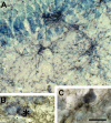
Synantocytes are closely associated with neurons. NG2-immunolabelled sections of adult rat brain counterstained with toluidine blue. (A) Synantocytes in the CA1 area of the hippocampus, some of which are directly apposed to pyramidal cell bodies, and extending processes through multiple layers. (B) Synantocyte processes forming a perineuronal network and enwrapping hippocampal neurons (asterisk). (C) Synantocyte apposed to and forming multiple contacts with a cortical pyramidal neuron. Scale bar = 25 µm.
Fig. 3
Fig. 3
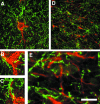
Synantocytes form multiple contacts with neurons and astrocytes. Confocal micrographs of cortex (A–C) and optic nerve (D,E) double immunofluorescence labelled for NG2 (green) and calbindin for neurons (red, A–C) or GFAP for astrocytes (red, D,E). Individual synantocytes form multiple contacts with neuronal somata (A), axons (B) and dendrites (C), and neurons are contacted by multiple synantocytes. In white matter, synantocytes are interspersed with astrocytes, which they contact (D), and their processes are interwined (E). Scale bar = 50 µm in A and D, and 12.5 µm in B, C and E.
Fig. 4
Fig. 4
Synantocytes contact nodes of Ranvier. Confocal micrographs of whole-mounted anterior medullary velum triple immunofluoresecnce labelling for NG2 (green), myelin basic protein (blue) and ankyrin-3G (red). (A) Individual synantocytes contact multiple nodes of Ranvier (curved arrows) and their processes closely follow the path of myelinated axons (inset). All synantocytes observed in the velum formed similar associations with nodes of Ranvier. (B) Deconvolution shows the synantocyte process extending along the myelin sheath to form exquisite contacts with the paranodes and node of Ranvier. Scale bar = 10 µm in A, 30 µm in inset and 50 µm in B.
Fig. 5
Fig. 5
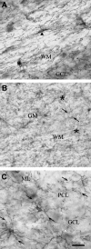
Synantocytes are interlaminar. NG2 immunolabelled sections of cerebellum (A, C) and cortex (B). (A) Individual cerebellar synantocytes with cell bodies in the white matter (WM, arrowhead) or at the interface between white and grey matter (GM, asterisk) extend processes into both. (B) Synantocytes in the cortical grey matter and subcortical white matter (asterisks) extend processes that traverse both layers and intermingle at the interface. (C) Interlaminar synantocytes extend processes into all layers of the cerebellum (arrows). Synantocytes are not specialized for either white or grey mater, and the same cells subserve both. Scale bar = 30 µm in A, C and 50 µm in B.
Fig. 6
Fig. 6
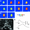
Synantocytes in vitro respond to glutamate and ATP with raised intracellular calcium. Explants of optic nerve glia were loaded with the calcium-sensitive dye fura-2 and imaged during bath application of ATP (A) or glutamate (B), and cells were identified at the end of the experiment by immunolabelling for NG2 (C). ATP and glutamate evoked a rapid increase in cytosolic [Ca2+]i in immunohistochemically identified synantocytes. (D) The response to ATP was transient and began to decay during exposure to the agonist, whereas the response to glutamate was slower to peak and was sustained after washout of the agonist.
Fig. 7
Fig. 7
Calcium signalling in synantocytes in situ. Optic nerves were isolated intact and loaded with fura-2 for calcium imaging, and at the end of the experiment cells were identified by immunolabelling for NG2 (A). Both ATP and glutamate (Glu) evoked a rise in cytosolic [Ca2+]i in immunohistochemically identified synantocytes, although the response to ATP was rapid and transient, whereas that to glutamate was slow and sustained (A). These responses were analysed in greater detail in unidentified glia, but all cells in the optic nerve responded similarly (n > 100), and it reasonable to conclude that the findings reflect synantocytes as well as astrocytes and oligodendrocytes. (B) The response to glutamate was markedly increased by incubation with cyclothiazide (CTZ), which acts on AMPA glutamate receptors to maintain them in an open state, and was blocked by the AMPA receptor antagonists NBQX. (C) The ATP response was blocked by suramin, a general antagonist for P2X and P2Y purinoceptors. (D) Suramin also inhibited the response to Glu plus CTZ, indicating the increase in [Ca2+]i was partly mediated by ATP released in response to activation of AMPA receptors. (E) Electrical stimulation of the optic nerve at 20 Hz for 20 s induced an increase in glial [Ca2+]i that was inhibited by suramin but not by NBQX. The results indicate that glial calcium signals are evoked by ATP, presumably released from astrocytes in response to axonal electrical activity and activation of AMPA glutamate receptors, and support a primary role for ATP as a ‘gliotransmitter’ in the optic nerve.

Similar articles

Cited by

References

    1. Aguirre A, Chittajallu R, Belachew S, Gallo V. NG2-expressing cells in the subventricular zone are type C-like cells that contribute to interneuron generation in the postnatal hippocampus. J Cell Biol. 2004;165:575–589. - PMC - PubMed
    1. Aguirre A, Gallo V. Postnatal neurogenesis and gliogenesis in the olfactory bulb from NG2-expressing progenitors of the subventricular zone. J Neurosci. 2004;24:10530–10541. - PMC - PubMed
    1. Araque A, Parpura V, Sanzgiri RP, Haydon PG. Tripartite synapses: glia, the unacknowledged partner. Trends Neurosci. 1999;22:208–215. - PubMed
    1. Barres BA, Koroshetz WJ, Swartz KJ, Chun LL, Corey DP. Ion channel expression by white matter glia: the O-2A glial progenitor cell. Neuron. 1990;4:507–524. - PubMed
    1. Belachew S, Chittajallu R, Aguirre AA, et al. Postnatal NG2 proteoglycan-expressing progenitor cells are intrinsically multipotent and generate functional neurons. J Cell Biol. 2003;161:169–186. - PMC - PubMed

Publication types

LinkOut - more resources