

Review
doi: 10.1111/j.1469-7580.2005.00452.x.
NG2: a component of the glial scar that inhibits axon growth
Affiliations
- PMID: 16367799
- PMCID: PMC1571583
- DOI: 10.1111/j.1469-7580.2005.00452.x
Item in Clipboard
Review
NG2: a component of the glial scar that inhibits axon growth
J Anat.
2005 Dec.
Abstract
NG2 is a high-molecular-weight chondroitin sulphate proteoglycan found on the surfaces of oligodendrocyte precursor cells (OPCs). Here we review the history and biology of OPCs with an emphasis on their functions after experimentally induced CNS injury. Injury to brain or spinal cord results in the rapid accumulation of NG2-expressing OPCs in the glial scar that forms at the injury site. The glial scar is considered a biochemical and physical barrier to successful axon regeneration and the functional properties of NG2 suggest that it, along with other macromolecules, participates in the creation of this growth-inhibitory environment. NG2 is an important target for therapies designed to promote successful axon regrowth.
Figures
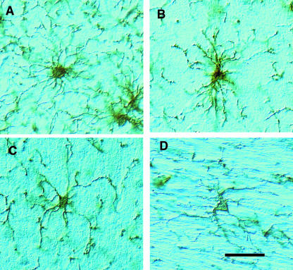
The appearance of NG2-labelled OPCs in the adult rat brain. Free-floating 30-µm-thick sections of adult rat brain were immunocytochemically stained with rabbit anti-NG2 antibodies. The individual cells shown are from (A) cortex, (B) suprapyramidal blade of the hippocampus, (C) external granule layer of cerebellum and (D) corpus callosum. The cells have irregularly shaped cell bodies from which numerous highly branched processes extend. The pattern of these processes is highly variable, being roughly spherical in the cortex but orientated parallel to axon bundles in the corpus callosum. The major processes give rise to many small filopodia. Scale bar = 50 µm.
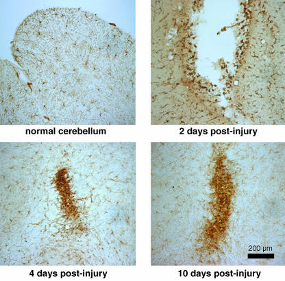
NG2-positive OPCs react to injury. Small stab wounds were made into the cerebellum of adult rats and the tissue was stained with anti-NG2 antibodies at the indicated survival times. By 2 days post-injury, many small intensely positive cells appear along the walls of the lesion cavity. At 4 and 10 days post-injury, the NG2-expressiong cells have formed small plaques at the injury site. These plaques correspond to the glial scar.

Adult DRGs grow poorly on beds of NG2-expressing Schwann cells. Left panel: normal adult rat sciatic nerve Schwann cells were maintained in culture and transfected with a full-length NG2 cDNA under control of the CMV promoter (pcDNA3.1). Both populations of Schwann cells express p75, the low-affinity neurotrophin receptor, but only the transfected cells express immunodetectable NG2. Right panel: adult rat DRG neurons were plated on the living Schwann cells and allowed to grow for 40 h and then stained with anti-βIII tubulin antibodies. Normal Schwann cells support extensive neurite growth: growth on the NG2-expressing Schwann cells was poor.
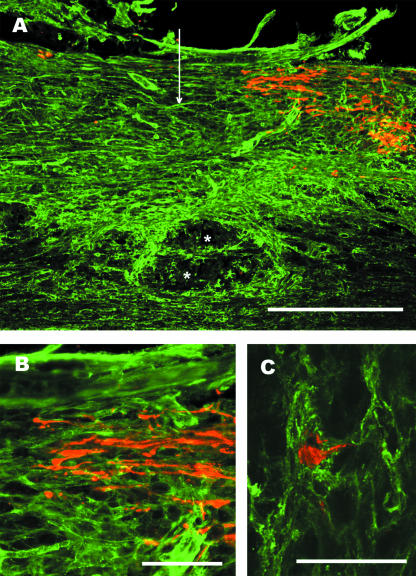
Lesioned mechanosensory axons end in an NG2-rich region of the glial scar. Adult rats were subjected to dorsal-over hemisections of the thoracic spinal cord and ascending mechanosensory axons retrogradely labelled by the injection of cholera toxin B subunit into the sciatic nerve 5 days post-lesion. Two days later the animals were killed and the tissue prepared for anti-NG2 (green) and anti-cholera toxin (red) immunofluorescence labelling. (A) NG2 immunoreactivity is increased surrounding and within the lesion centre. The vertical arrow marks the lesion centre and the asterisks indicate a cyst forming near the central canal. Scale bar = 500 µm. (B) Higher power view showing that the retrogradely labelled axons end with dystrophic end bulbs in a area with dense anti-NG2 stain. Scale bar = 100 µm. (C) A dystrophic end bulb is encased in NG2-positive processes. This is a z-stack made from 18 1-µm-thick scans taken with a Ziess 510 confocal microscope. Scale bar = 50 µm.
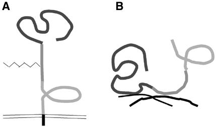
NG2 can adapt in different conformations. These schematics illustrate a model for the different conformations of NG2. The three individual domains of the ECD are indicated by the different shades of grey. (A) As an integral membrane proteoglycan, NG2 may extend away from the membrane surface. In this conformation, domain 1 is available but domain 3 may be shielded and inaccessible to neural growth cones. (B) When shed or secreted from the membrane, NG2 can become incorporated into the extracellular matrix via the collagen binding domain 2. Both domains 1 and 3 are now accessible. Because both domains 1 and 3 can independently inhibit neurite growth, extracellular NG2 may amplify the growth-non-permissive properties of the glial scar.
Similar articles
-
Inhibition of axon growth by oligodendrocyte precursor cells.Mol Cell Neurosci. 2002 May;20(1):125-39. doi: 10.1006/mcne.2002.1102. Mol Cell Neurosci. 2002. PMID: 12056844
-
Oligodendrocyte precursor cells: reactive cells that inhibit axon growth and regeneration.J Neurocytol. 2002 Jul-Aug;31(6-7):481-95. doi: 10.1023/a:1025791614468. J Neurocytol. 2002. PMID: 14501218 Review.
-
Evidence that perinatal and adult NG2-glia are not conventional oligodendrocyte progenitors and do not depend on axons for their survival.Mol Cell Neurosci. 2003 Aug;23(4):544-58. doi: 10.1016/s1044-7431(03)00176-3. Mol Cell Neurosci. 2003. PMID: 12932436
-
Mixed primary culture and clonal analysis provide evidence that NG2 proteoglycan-expressing cells after spinal cord injury are glial progenitors.Dev Neurobiol. 2007 Jun;67(7):860-74. doi: 10.1002/dneu.20369. Dev Neurobiol. 2007. PMID: 17506499
-
Polydendrocytes: NG2 cells with many roles in development and repair of the CNS.Neuroscientist. 2007 Feb;13(1):62-76. doi: 10.1177/1073858406295586. Neuroscientist. 2007. PMID: 17229976 Review.
Cited by
-
Role of the lesion scar in the response to damage and repair of the central nervous system.Cell Tissue Res. 2012 Jul;349(1):169-80. doi: 10.1007/s00441-012-1336-5. Epub 2012 Feb 25. Cell Tissue Res. 2012. PMID: 22362507 Free PMC article. Review.
-
The Significance of Chondroitin Sulfate Proteoglycan 4 (CSPG4) in Human Gliomas.Int J Mol Sci. 2018 Sep 12;19(9):2724. doi: 10.3390/ijms19092724. Int J Mol Sci. 2018. PMID: 30213051 Free PMC article. Review.
-
Understanding the Role of the Glial Scar through the Depletion of Glial Cells after Spinal Cord Injury.Cells. 2023 Jul 13;12(14):1842. doi: 10.3390/cells12141842. Cells. 2023. PMID: 37508505 Free PMC article. Review.
-
Glycogen synthase kinase 3 beta (GSK3β) at the tip of neuronal development and regeneration.Mol Neurobiol. 2014 Apr;49(2):931-44. doi: 10.1007/s12035-013-8571-y. Epub 2013 Oct 25. Mol Neurobiol. 2014. PMID: 24158777 Review.
-
Morphological and physiological interactions of NG2-glia with astrocytes and neurons.J Anat. 2007 Jun;210(6):661-70. doi: 10.1111/j.1469-7580.2007.00729.x. Epub 2007 Apr 25. J Anat. 2007. PMID: 17459143 Free PMC article. Review.
References
-
- Bradbury EJ, Moon LD, Popat RJ, et al. Chondroitinase ABC promotes functional recovery after spinal cord injury. Nature. 2002;416:636–640. - PubMed
-
- Bu J, Akhtar N, Nishiyama A. Transient expression of the NG2 proteoglycan by a sub-population of activated macrophages in an excitotoxic hippocampal lesion. Glia. 2001;34:296–310. - PubMed
-
- Butt AM, Duncan A, Hornby MF, et al. Cells expressing the NG2 antigen contact nodes of Ranvier in adult CNS white matter. Glia. 1999;26:84–91. - PubMed
-
- Butt AM, Kiff J, Hubbard P, Berry M. Synaptocyte: new functions for novel NG2 expressing glia. J Neurocytol. 2002;31:551–565. - PubMed
Publication types
MeSH terms
Substances
LinkOut - more resources
Full Text Sources
Other Literature Sources
Medical
