. 2005 Dec;207(6):745-61.
doi: 10.1111/j.1469-7580.2005.00491.x.

Characterization of the rhesus monkey superior olivary complex by calcium binding proteins and synaptophysin

Affiliations

Characterization of the rhesus monkey superior olivary complex by calcium binding proteins and synaptophysin

Ivonne Bazwinsky et al. J Anat. 2005 Dec.

Abstract

This study was performed in order to characterize the main nuclei of the rhesus monkey superior olivary complex by means of antibodies against the calcium binding proteins parvalbumin, calbindin and calretinin and the synaptic vesicle protein synaptophysin. These markers revealed the neuronal morphology and organization of nuclei located within the rhesus monkey superior olivary complex. The architectural details included the distribution of axonal terminals on neurons. The medial superior olivary nucleus was present as a column of neurons. No clear segregation of calretinin-positive terminals was noticed on the medial and lateral dendritic fields of these neurons. The lateral superior olivary nucleus was characterized by a distinct nuclear shape. Calretinin-, parvalbumin- or calbindin-positive terminals contacted somata and dendrites. The medial nucleus of trapezoid body could be clearly differentiated as a distinct region in the rhesus monkey superior olivary complex. Somata of that nucleus showed calbindin- and parvalbumin-labelling whereas somatic calyces of Held were reavealed by calretinin and synaptophysin labelling. The results are discussed with respect to the processing of acoustic information in primate species and their ability to hear high and low frequencies, which is reflected by anatomical correlates.


Figures
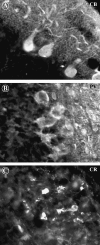
Fig. 1
Fig. 1
(A,B) Purkinje cells express calbindin (CB) and parvalbumin (PV), but no calretinin (CR). (C) CR is solely seen in granule cells of the granule cell layer.
Fig. 2
Fig. 2
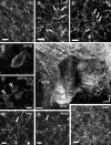
Overview (E) and details (A–D and F–I) of SYN-labelled sections of the superior olivary complex. Sections are in the transverse plane. (A) Numerous SYN-positive terminals are found in the LSO area. (B) Arrows indicate SYN-labelled terminals, which appeared in apposition to the somatic membrane of an LSO neuron. (C) The packing density of SYN-labelled terminals visualized the contours of dendrites of LSO neurons (arrows) in some cases. (D) High-magnification view of an MNTB soma, which is strongly enclosed by SYN-labelled synapses. (F) Open triangles point to somata covered by SYN-immunoreactive bouton-like terminals. (G,H) SYN-positive terminals (arrows) distributed on somata and distal dendrites of MSO neurons. (I) Heavily labelled and tightly localized SYN-positive terminals in the MPO. Scale bars: A–D, F–I = 15 µm, E = 200 µm.
Fig. 3
Fig. 3
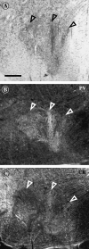
Transverse sections of rhesus monkey brainstem through the SOC. (A) Nissl-stained sections, which show LSO, MSO and the MNTB (triangles). (B,C) PV- and CR-labelled brainstem sections of the rhesus monkey SOC-nuclei MNTB, LSO and MSO (triangles) from the same level as shown in A. Scale bar = 500 µm.
Fig. 4
Fig. 4
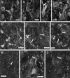
Details of PV (A–D,J)-, CR (E–H)- and CB (I,K)-labelling as seen with confocal laser scanning microscopy in MSO. (A–D) MSO neurons (open triangles) show weak and strong PV-labelling. Additionally, strongly PV-labelled terminals (arrows) were found on dendrites (A,C) and somata (B,D). (E) A restricted number of MSO neurons were immunoreactive for CR. Arrowheads point to CR-positive dendrites of bipolar neurons. (F) Large CR-positive synaptic endings (arrows, inset) contact the somata and proximal dendrites of MSO neurons. (G) CR-immunoreactive terminals (arrow, inset) also found on distal dendrites of MSO neurons. (H) CR-positive synaptic endings (arrows) also terminate on somata and dendrites of CR-immunonegative neurons. (I) CB-immunoreactivity is restricted to small terminals and axonal swellings (arrows). (J,K) Arrow in J points to a PV-positive somatic terminal, which is also CB-immunoreactive as seen in K (arrow). Scale bars: A,E,I = 30 µm, B–D,G,J,K = 15 µm, F,H = 10 µm.
Fig. 5
Fig. 5
Details of PV (A–D,I)-, CR (E–G)- and CB (H,J)-labelling as seen with confocal laser scanning microscopy in LSO. (A–D) Most LSO neurons express PV. Neurons have round, bipolar or multipolar shaped somata. Sometimes PV-positive terminals are seen on somata and dendrites of these cells (arrows). (E) Strongly and weakly labelled LSO neurons (open triangles) embedded in CR-positive neuropil. (F,G) Numerous CR-positive terminals cover somata and distal dendrites of LSO neurons (arrows). (H) Many CB-positive fibres (arrow) and small synaptic endings located within the LSO. (I,J) Arrows point to terminals on a single LSO neuron, which is PV-positive as shown in I. Note the same neuron contains no immunoreactivity for CB (J) whereas some of the terminals coexpress PV and CB. Scale bars: B–D,F = 15 µm, A,E,H = 30 µm, G = 10 µm, I = 15 µm.
Fig. 6
Fig. 6
Details of PV (A–D)-, CR (E–H)- and CB (I–M)-labelling as seen with confocal laser scanning microscopy in MNTB. (A–D) MNTB neurons are immunoreactive for PV. Neurons are characterized by multipolar (A) or the typical globular soma shape (B). PV-positive terminals (arrows) contact somata and dendrites of MNTB neurons (B). (E) CR-immunoreactivity visualizes calyces of Held, whereas somata are CR-negative (open triangles). (F,G) Thick axonal stump bifurcates (arrows) and forms finger-like endings, which strongly associated with CR-negative somata. (H) Such a calyx of Held (arrow) encloses the whole cell soma of an MNTB neuron. (I) MNTB neurons in the lateral part of MNTB show the strongest CB-labelling intensity (open triangle). CB-labelling reaches into the distal dendrites (arrowhead in the inset). (J,K) The heavily CB-labelled somata of MNTB neurons form bushy-like dendritic trees (arrowheads). (L,M) Two coronal sections through MNTB showing that densely packed CB-labelled somata were more concentrated in the lateral part than in its central and medial aspects. Scale bars: D = 10 µm, A–C,F–H,J,K = 15 µm, I = 30 µm, E = 50 µm.

Similar articles

Cited by

References

    1. Adams JC, Mugnaini E. Immunocytochemical evidence for inhibitory and disinhibitory circuits in the superior olive. Hear Res. 1990;49:281–298. - PubMed
    1. Andressen C, Blümcke I, Celio MR. Calcium-binding proteins: selective markers of nerve cells. Cell Tiss Res. 1993;271:181–208. - PubMed
    1. Baimbridge KG, Celio MR, Rogers JH. Calcium binding proteins in the nervous system. Trends Neurosci. 1992;15:303–308. - PubMed
    1. Banks MI, Smith PH. Intracellular recordings from neurobiotin-labeled cells in brain slices of the rat medial nucleus of the trapezoid body. J Neurosi. 1992;12:2819–2837. - PMC - PubMed
    1. Bazwinsky I, Haertig W, Rübsamen R. Distribution of different calcium binding proteins in the cochlear nucleus and superior olivary complex of gerbils and opossum. Abstract ARO Midwinter Mtg. 1999;22:260.

LinkOut - more resources