

Changes in adipocytes and dendritic cells in lymph node containing adipose depots during and after many weeks of mild inflammation
- PMID: 16367804
- PMCID: PMC1571578
- DOI: 10.1111/j.1469-7580.2005.00506.x
Changes in adipocytes and dendritic cells in lymph node containing adipose depots during and after many weeks of mild inflammation
Abstract
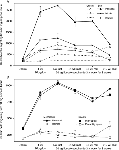
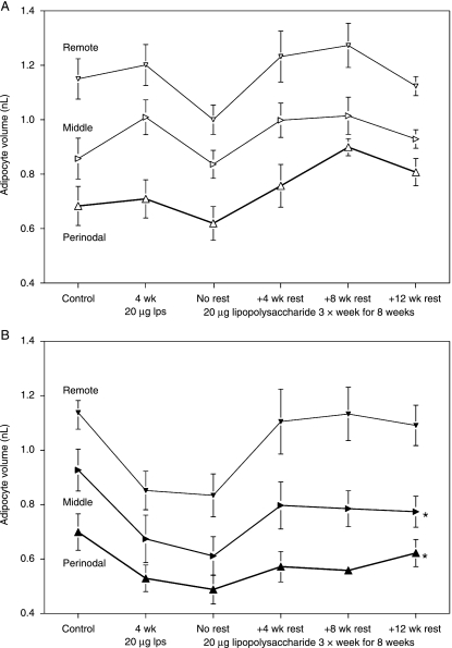
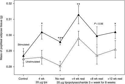
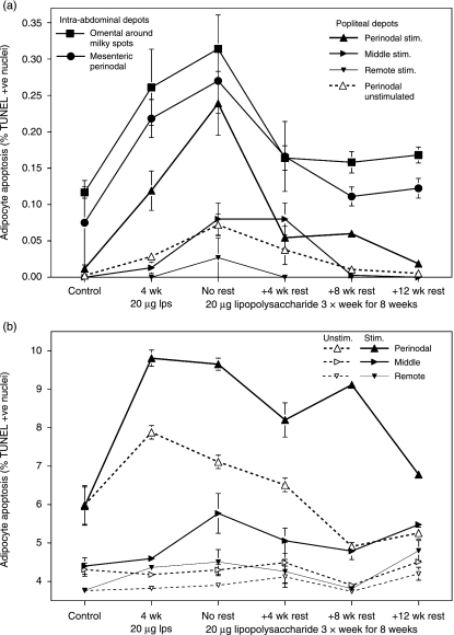
The time course and cellular basis for inflammation-induced hypertrophy of adipose tissue were investigated over 20 weeks in mature male rats. Mild inflammation was induced by subcutaneous injection of 20 microg lipopolysaccharide into one hind-leg three times/week for 4 or 8 weeks, followed by up to 12 weeks 'rest' without intervention. Mean volume and frequency of apoptosis (TUNEL assay) were measured in adipocytes isolated from sites defined by their anatomical relations to lymph nodes, plus numbers of CCL21-stimulated lymph node-derived and adipose tissue-derived dendritic cells. Experimental inflammation increased dendritic cells and adipocyte apoptosis in the locally stimulated popliteal depot and the lymphoid tissue-associated regions of the contralateral popliteal and mesentery and omentum. Responses declined slowly after inflammation ended, but all measurements from the locally stimulated popliteal depot, and the omentum, were still significantly different from controls after 12 weeks rest. The locally stimulated popliteal adipose tissue enlarged by 5% within 4 weeks and remained larger than the control. We conclude that prolonged inflammation induces permanent enlargement, greater adipocyte turnover and increased dendritic cell surveillance in the adjacent adipose tissue and the omentum. The experiment suggests a mechanism for selective hypertrophy of lymphoid tissue-associated adipose tissue in chronic stress and inflammatory disorders, including impaired lymph drainage, Crohn's disease and HIV-associated lipodystrophy, and a link between evolutionary fitness, sexual selection and aesthetically pleasing body symmetry. It would be useful for further study of molecular mechanisms in inflammation-induced local hypertrophy of adipose tissue and development of specific therapies that avoid interference with whole-body lipid metabolism.
Figures
References
-
- Björntorp P. The regulation of adipose-tissue distribution in humans. Int J Obesity. 1996;20:291–302. - PubMed
-
- Curat CA, Miranville A, Sengenès C, et al. From blood monocytes to adipose tissue-resident macrophages – Induction of diapedesis by human mature adipocytes. Diabetes. 2004;53:1285–1292. - PubMed
-
- Dhurandhar NV, Kulkarni PR, Ajinkya SM, Sherikar AA, Atkinson RL. Association of adenovirus infection with human obesity. Obes Res. 1997;5:464–469. - PubMed
-
- Dhurandhar NV, Israel BA, Kolesar JM, Mayhew GF, Cook ME, Atkinson RL. Increased adiposity in animals due to a human virus. Int J Obesity. 2000;24:989–996. - PubMed
MeSH terms
Substances
LinkOut - more resources
Full Text Sources
