. 2006 Jan;8(1):107-19.
doi: 10.1111/j.1462-5822.2005.00604.x.

Cytolysin-dependent delay of vacuole maturation in macrophages infected with Listeria monocytogenes

Affiliations

Cytolysin-dependent delay of vacuole maturation in macrophages infected with Listeria monocytogenes

Rebecca Henry et al. Cell Microbiol. 2006 Jan.

Abstract

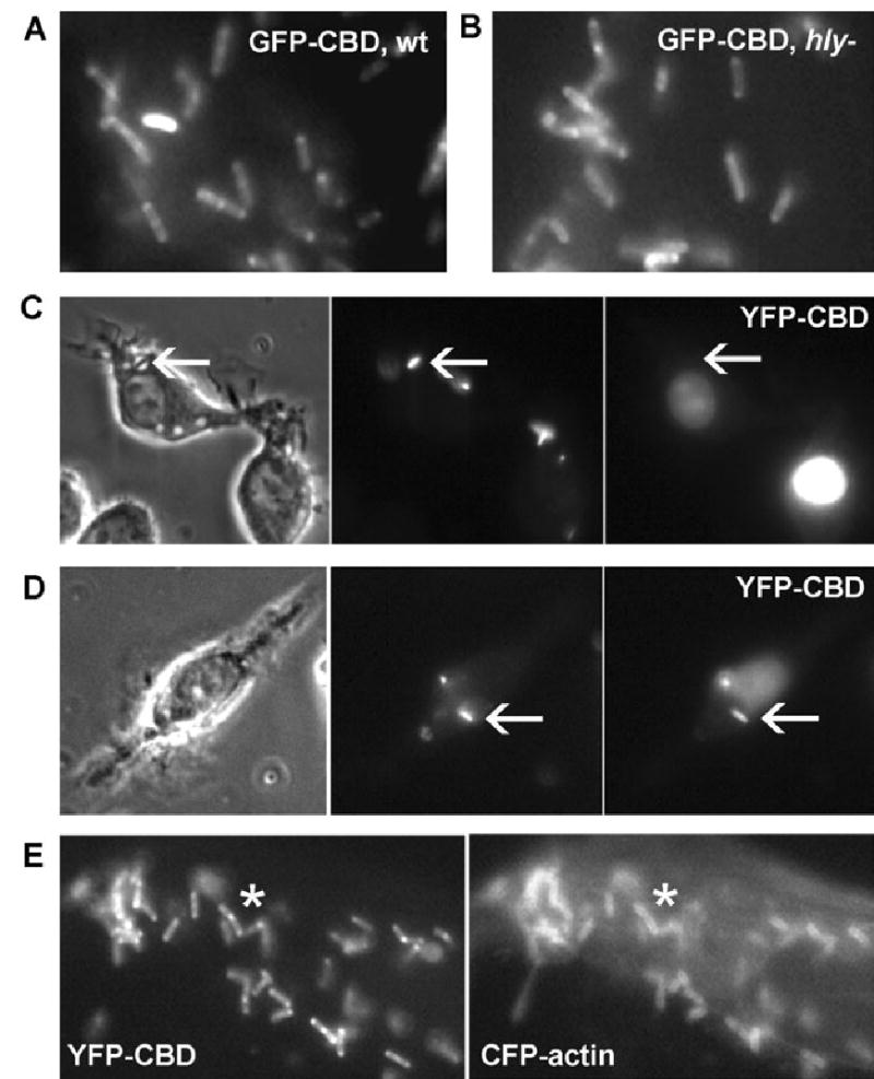
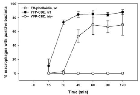
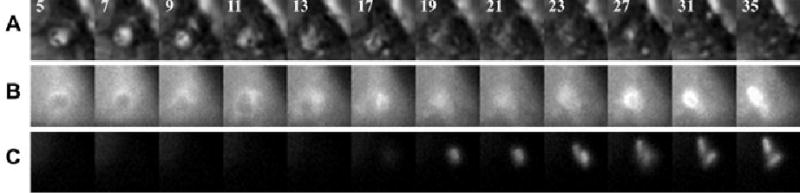
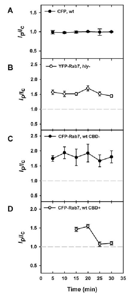
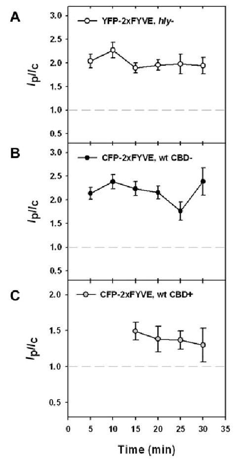
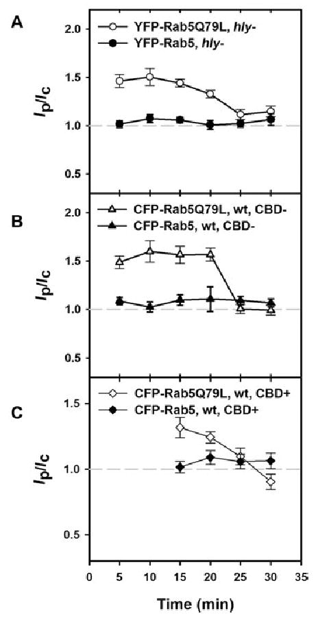
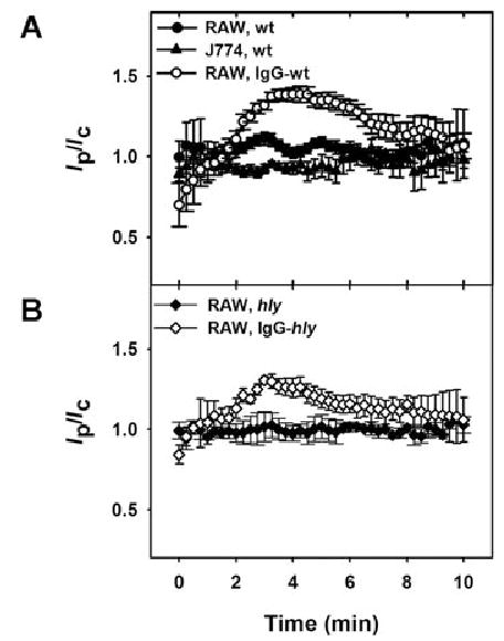
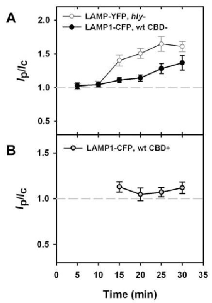
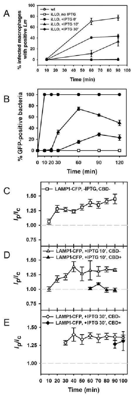
The bacterial pathogen Listeria monocytogenes (Lm) evades the antimicrobial mechanisms of macrophages by escaping from vacuoles to the cytosol, through the action of the cytolysin listeriolysin O (LLO). Because of heterogeneities in the timing and efficiency of escape, important questions about the contributions of LLO to Lm vacuole identity and trafficking have been inaccessible. Expression of cyan fluorescent protein (CFP)-labelled endocytic membrane markers in macrophages along with a yellow fluorescent protein (YFP)-labelled indicator of Lm entry to the cytosol identified compartments lysed by bacteria. Lm escaped from Rab5a-negative, lysosome-associated membrane protein-1 (LAMP1)-negative, Rab7-positive, phosphatidylinositol 3-phosphate [PI(3)P]-positive vacuoles. Lm vacuoles did not label with YFP-Rab5a unless the bacteria were first opsonized with IgG. Wild-type Lm delayed vacuole fusion with LAMP1-positive lysosomes, relative to LLO-deficient Lm. Bacteria prevented from expressing LLO until their arrival into LAMP1-positive lysosomes escaped inefficiently. Thus, the LLO-dependent delay of Lm vacuole fusion with lysosomes affords Lm a competitive edge against macrophage defences by providing bacteria more time in organelles they can penetrate.


Figures

Fig. 1
Fig. 1
YFP-CBD as a marker of Lm delivery into cytosol. A and B. Epifluorescence images of (A) wild-type Lm and (B) hly Lm decorated with HGFP-CBD after incubation with the purified fluorescent protein. C and D. Phase-contrast (left), SNARF-labelled Lm (middle) and YFP-CBD fluorescence (right) images. (C) Cells viewed at 10 min after infection contained bacteria in phase-bright vacuoles (arrows) which were not labelled with YFP-CBD. (D) Cells viewed 60 min after infection showed some bacteria labelled with YFP-CBD (arrows). E. YFP fluorescence image of a RAW 264.7 macrophage showing YFP-CBD-positive cytosolic Lm (left) with corresponding labelling by CFP-actin (asterisks).
Fig. 2
Fig. 2
YFP-CBD marked cytosolic Lm within 15 min of infection. Macrophages were infected with wild-type or hly Lm for 3 min and washed, resulting in <1 bacterium per macrophage. Infected cells were incubated further and fixed at the indicated time points. Bacteria were identified by DAPI staining; cytosolic bacteria were identified by their labelling with either YFP-CBD or TR-phalloidin. At each time point >200 infected cells were scored for the presence of YFP-CBD or TR-phalloidin. YFP-CBD labelled wild-type Lm (black circles) between 15 and 30 min, while TR-phalloidin stained wild-type Lm (grey circles) 45–120 min after infection. YFP-CBD did not decorate hly Lm (open circles) at any time. Each data point represents the mean ± standard error of the mean (SEM) of four experiments.
Fig. 3
Fig. 3
YFP-CBD allowed identification of compartments lysed by Lm. Macrophages were co-transfected with YFP-CBD and CFP-Rab7. Cells were infected with wild-type Lm, then phase-contrast, YFP and CFP images of infected cells were taken at 2 min intervals. Each column contains component images of one time point, indicated in (A) as the time, in minutes, after the start of infection. A. Phase-contrast images show two bacteria in a phase-bright vacuole between 5 and 17 min, after which the vacuole became phase-dark. B. CFP-Rab7 labelling of this vacuole illustrated that the vacuole collapsed after 17 min, but fluorescence persisted. CFP fluorescence appeared to increase over time due to movement of the vacuole within the focal plane. C. Coincident with collapse of the vacuole, the bacteria acquired YFP-CBD. From these images it was apparent that bacteria were present in a Rab7-positive compartment during escape to the cytosol.
Fig. 4
Fig. 4
Lm lysed compartments labelled with Rab7. The average fluorescence intensity of the phagosome (Ip) was divided by the average fluorescence intensity of the entire cell (Ic) to produce the cell-normalized phagosome fluorescence (Ip/Ic). Each symbol represents the mean values of >45 vacuoles measured in multiple experiments. A. Ip/Ic ratios for Lm vacuoles in macrophages expressing CFP were near 1.0 from 5 to 30 min. These values were compared as a negative control to all further experiments (grey dotted line in subsequent panels). B and C. (B) hly Lm and (C) YFP-CBD-negative, wild-type Lm resided in Rab7-positive vacuoles between 5 and 30 min after infection, as indicated by Ip/Ic values significantly higher than those in cells expressing CFP (P < 0.01). One hundred per cent of Lm vacuoles were Rab7-positive. D. YFP-CBD-positive, wild-type Lm displayed Ip/Ic ratios greater than 1.5 for CFP-Rab7 from 15 to 20 min, demonstrating that Lm lysed compartments labelled with CFP-Rab7. The bars indicate SEM.
Fig. 5
Fig. 5
PI(3)P on vacuoles lysed by Lm. A and B. YFP- or CFP-2xFYVE-labelled vacuoles containing (A) hly Lm and (B) YFP-CBD-negative, wild-type Lm at 5–30 min. One hundred per cent of Lm vacuoles were 2xFYVE-positive. C. CFP-2xFYVE and YFP-CBD colocalized 15–20 min after infection, signifying the presence of PI(3)P on vacuoles perforated by wild-type bacteria. At each time point >45 vacuoles were measured. The bars indicate SEM.
Fig. 6
Fig. 6
Rab5 did not label Lm vacuoles, but localization of Rab5Q79L to Lm vacuoles did not prevent escape. A. While YFP-Rab5a did not localize to vacuoles containing hly Lm (closed circles), YFP-Rab5Q79L was present from 5 to 20 min (open circles). B. CFP-Rab5Q79L localized to CBD-negative, wild-type Lm vacuoles from 5 to 20 min (open triangles), but CFP-Rab5a did not (closed triangles). C. CFP-Rab5a was not associated with YFP-CBD-positive Lm at any time point measured (closed diamonds). CFP-Rab5Q79L associated with CBD-positive Lm at 15–20 min (open diamonds), demonstrating that Lm could lyse vacuoles labelled with Rab5Q79L. At each time point >45 vacuoles were measured. The bars indicate SEM.
Fig. 7
Fig. 7
Rab5a was not present on Lm vacuoles, but did localize transiently to vacuoles containing IgG-opsonized Lm. A. The internalization of wild-type Lm was observed in RAW 264.7 (closed circles) or J774 macrophages (closed triangles). YFP-Rab5a was not associated with wild-type Lm vacuoles during internalization or the first 5 min subsequent to internalization. YFP-Rab5a associated transiently with vacuoles during entry of IgG-opsonized Lm into RAW 264.7 macrophages (open circles). B. YFP-Rab5a did not associate with vacuoles containing hly Lm (closed diamonds), but did associate transiently with vacuoles containing IgG-opsonized, hly Lm (open diamonds). The data were aligned by the timing of phase-bright vacuole formation, which was set at 3 min. At each time point >15 vacuoles were measured. The bars indicate SEM.
Fig. 8
Fig. 8
Wild-type Lm phagosomes showed delayed acquisition of LAMP1-CFP. A. Vacuoles containing hly bacteria (open circles) acquired LAMP1-YFP from 15 to 30 min after infection, as indicated by Ip/Ic values near 1.5. Ninety per cent of vacuoles were LAMP1-positive at 15 min, and 100% of vacuoles were LAMP1-positive at 30 min. The acquisition of LAMP1-CFP by vacuoles containing CBD-negative wild-type Lm was delayed relative to hly phagosomes. Ip/Ic did not reach a value significantly higher than 1.0 until 25 min (P < 0.01). Three per cent of vacuoles containing CBD-negative wild-type Lm were LAMP1-positive at 15 min, and 96% were LAMP1-positive at 30 min. B. Lack of colocalization of LAMP1-CFP with YFP-CBD indicated that Lm escaped to the cytosol before association with LAMP1-CFP labelled lysosomes. While Ip/Ic ratios for CBD-negative, wild-type Lm were high from 25 to 30 min, Ip/Ic ratios for CBD-positive wild-type Lm remained low (P < 0.01). At each time point >45 vacuoles were measured. The bars indicate SEM.
Fig. 9
Fig. 9
Perforation of LAMP1-positive compartments by LLO was inefficient. A. Macrophages expressing YFP-CBD were infected with wild-type or iLLO Lm to produce <1 bacterium per macrophage and were fixed at the indicated time points. Infected cells were identified by DAPI staining and cytosolic bacteria were identified by YFP-CBD labelling. YFP-CBD labelled wild-type Lm (grey circles) by 60 min. Escape of iLLO Lm treated with 10 mM IPTG before infection (IPTG 0′, black circles) was reduced relative to wild type. iLLO Lm treated with IPTG at 10 min after infection (IPTG 10′, triangles) were present in the cytosol at 60 min. iLLO Lm treated with IPTG at 30 min (IPTG 30′, diamonds) did not become CBD-labelled until 90 min, at which point ~0.5% were YFP-CBD-positive. In the absence of IPTG (open squares, partially obscured by IPTG 30′ data), no Lm were CBD-labelled. At each time point >100 infected cells were scored for the presence of YFP-CBD. Each data point represents the mean ± SEM of two experiments. B. Overnight cultures of iGFP Lm or macrophages infected with iGFP Lm were fixed at the indicated time points. Intracellular bacteria were identified by DAPI staining and GFP-producing bacteria were identified by indirect immunofluorescence. iGFP Lm treated with IPTG in broth (black circles) stained positive for GFP by 10 min. iGFP Lm treated with IPTG at 10 min after infection stained positive for GFP from 30 to 120 min (black triangles), whereas iGFP Lm treated with IPTG at 30 min stained positive for GFP from 60 to 120 min (black diamonds). iGFP Lm did not stain positive for GFP in the absence of IPTG treatment (open squares). At each time point 100 bacteria were scored for Alexa Fluor 594 staining. Each data point represents the mean ± SEM of two experiments. C. Uninduced iLLO Lm acquired LAMP1-CFP by 20 min after infection, but did not decorate with YFP-CBD at any time (open squares). D. After treatment with IPTG at 10 min after infection, iLLO Lm escaped from LAMP1-CFP-negative compartments at 60–90 min (closed triangles). Many CBD-negative vacuoles acquired LAMP1-CFP as early as 20 min (open triangles). E. In cells treated with IPTG at 30 min after infection, iLLO Lm perforated LAMP1-CFP-positive vacuoles at 90–100 min (closed diamonds).

References

    1. Alvarez-Dominguez C, Stahl PD. Increased expression of Rab5a correlates directly with accelerated maturation of Listeria monocytogenes phagosomes. J Biol Chem. 1999;274:11459–11462. - PubMed
    1. Alvarez-Dominguez C, Roberts R, Stahl PD. Internalized Listeria monocytogenes modulates intracellular trafficking and delays maturation of the phagosome. J Cell Sci. 1997;110:731–743. - PubMed
    1. Beauregard KE, Lee KD, Collier RJ, Swanson JA. pH-dependent perforation of macrophage phagosomes by listeriolysin O from Listeria monocytogenes. J Exp Med. 1997;186:1159–1163. - PMC - PubMed
    1. de Chastellier C, Berche P. Fate of Listeria monocytogenes in murine macrophages: evidence for simultaneous killing and survival of intracellular bacteria. Infect Immun. 1994;62:543–553. - PMC - PubMed
    1. Conte MP, Petrone G, Longhi C, Valenti P, Morelli R, Superti F, Seganti L. The effects of inhibitors of vacuolar acidification on the release of Listeria monocytogenes from phagosomes of Caco-2 cells. J Med Microbiol. 1996;44:418–424. - PubMed

Publication types

MeSH terms

Substances