

Differential expression of S100A2 and S100A4 in lung adenocarcinomas: clinicopathological significance, relationship to p53 and identification of their target genes
- PMID: 16367903
- PMCID: PMC11159992
- DOI: 10.1111/j.1349-7006.2005.00121.x
Differential expression of S100A2 and S100A4 in lung adenocarcinomas: clinicopathological significance, relationship to p53 and identification of their target genes
Abstract
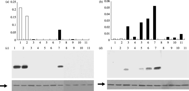
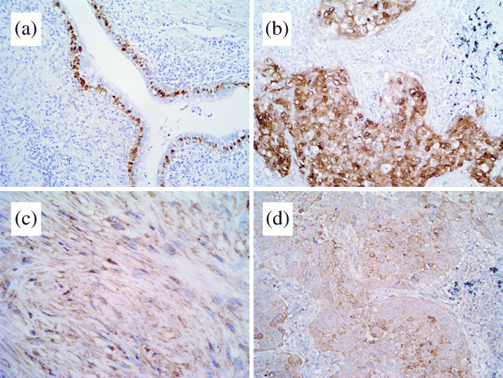
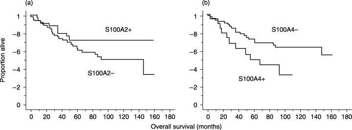
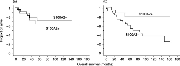
Previous studies suggest that some S100 proteins are involved in the progression of certain types of cancer. However, no comprehensive data is currently available on the expression of S100 family genes in lung adenocarcinomas. Oligonucleotide array, quantitative reverse transcription-polymerase chain reaction and western blot analyses of lung adenocarcinoma cell lines and bronchiolar epithelial cells (SAEC and NHBE) revealed that S100A2 and S100A4 were the most strikingly downregulated and upregulated members of the S100 family, respectively. Immunohistochemical analyses of 94 primary lung adenocarcinomas showed that positive S100A2 expression (33/94, 35.1%) was significantly associated with lymphatic invasion (P=0.0233) and positive S100A4 expression (19/94, 20.2%) with vascular invasion (P=0.0454). Interestingly, a strong inverse relationship was found between S100A4 and p53 expression (P=0.0008). Survival analyses showed that S100A4 positivity was associated with poor patient prognosis (P=0.042). S100A2 positivity was not associated with patient survival when the whole patient group was analyzed; however, S100A2 positivity was a favorable prognostic indicator in patients with p53-negative tumors (P=0.0448). Finally, we used oligonucleotide array analyses and identified potential S100A2 and S100A4 target genes involved in cancer progression: S100A2 induced RUNX3 and REPRIMO; S100A4 induced EZRIN, RUNX1 and WISP1; S100A2 repressed EGFR, NFKB2 and RELA2; and S100A4 repressed ANXA10 and IL1RN. Thus, the present study demonstrates involvement of S100A2 and S100A4 in the progression of lung adenocarcinomas and an inverse association between S100A4 and p53 expression, and provides a list of targets regulated by S100A2 and S100A4.
(Cancer Sci 2005; 96: 844 - 857).
Figures
References
-
- Statistics and Information Department. Vital Statistics, 2000. Tokyo: Ministry of Health, Labor and Welfare, 2001.
-
- Janssen‐Heijnen ML, Coebergh JW. Trends in incidence and prognosis of the histological subtypes of lung cancer in North America, Australia, New Zealand and Europe. Lung Cancer 2001; 31: 123–37. - PubMed
-
- Naruke T, Tsuchiya R, Kondo H, Asamura H, Nakayama H. Implications of staging in lung cancer. Chest 1997; 112: 242S–8S. - PubMed
-
- Moriya Y, Niki T, Yamada T, Matsuno Y, Kondo H, Hirohashi S. Increased expression of laminin‐5 and its prognostic significance in lung adenocarcinomas of small size. An immunohistochemical analysis of 102 cases. Cancer 2001; 91: 1129–41. - PubMed
-
- Danesi R, De Braud F, Fogli S et al. Pharmacogenetics of anticancer drug sensitivity in non‐small cell lung cancer. Pharmacol Rev 2003; 55: 57–103. - PubMed
MeSH terms
Substances
LinkOut - more resources
Full Text Sources
Other Literature Sources
Medical
Research Materials
Miscellaneous
