. 2006 Jan;143(1):15-23.
doi: 10.1111/j.1365-2249.2005.02958.x.

Inhibition of classical complement activation attenuates liver ischaemia and reperfusion injury in a rat model

Affiliations

Inhibition of classical complement activation attenuates liver ischaemia and reperfusion injury in a rat model

B H M Heijnen et al. Clin Exp Immunol. 2006 Jan.

Abstract

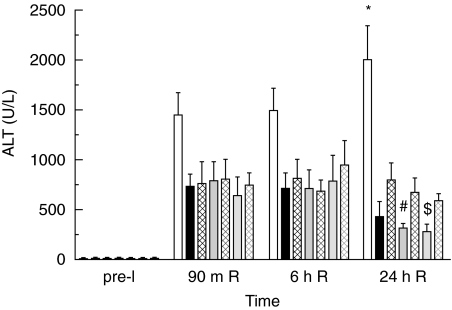
Activation of the complement system contributes to the pathogenesis of ischaemia/reperfusion (I/R) injury. We evaluated inhibition of the classical pathway of complement using C1-inhibitor (C1-inh) in a model of 70% partial liver I/R injury in male Wistar rats (n = 35). C1-inh was administered at 100, 200 or 400 IU/kg bodyweight, 5 min before 60 min ischaemia (pre-I) or 5 min before 24 h reperfusion (end-I). One hundred IU/kg bodyweight significantly reduced the increase of plasma levels of activated C4 as compared to albumin-treated control rats and attenuated the increase of alanine aminotransferase (ALT). These effects were not better with higher doses of C1-inh. Administration of C1-inh pre-I resulted in lower ALT levels and higher bile secretion after 24 h of reperfusion than administration at end-I. Immunohistochemical assessment indicated that activated C3, the membrane attack complex C5b9 and C-reactive protein (CRP) colocalized in hepatocytes within midzonal areas, suggesting CRP is a mediator of I/R-induced, classical complement activation in rats. Pre-ischaemic administration of C1-inh is an effective pharmacological intervention to protect against liver I/R injury.


Figures

Fig. 1
Fig. 1
Bile secretion (means ± SEM) measured during 15 min after 24 h of reperfusion (24 h R). Rats were treated with albumin (white bar), 100 IU/kg C1-inh pre-I (black bar), 100 IU/kg C1-inh end-I (black chequered bar), 200 IU/kg C1-inh pre-I (dark grey bar), 200 IU/kg C1-inh end-I (dark grey chequered bar), 400 IU/kg C1-inh pre-I (light grey bar) and 400 IU/kg C1-inh end-I (light grey chequered bar). All rats treated with C1-inh pre-I, and rats treated with 400 IU/kg of C1-inh end-I, showed higher bile secretion than albumin treated rats. *significant versus albumin treated rats, P < 0·01. Rats treated with 200 IU/kg C1-inh pre-I showed better bile secretion than rats treated with the same dose at the end of ischaemia. ♯significant versus 200 IU pre-ischaemia, P < 0·05. Previous studies showed normal baseline levels of bile production in nonischaemic rats of 0·018 ml/min [32].
Fig. 2
Fig. 2
Plasma ALT (means ± SEM) measured pre-ischaemia (pre-I), after 90 m of reperfusion (90 m R), 6 h of reperfusion (6 h R) and 24 h of reperfusion (24 h R) in rats receiving albumin or C1-inh. After 24 h R, ALT levels in C1-inh treated rats were significantly lower than in albumin-treated rats (*P < 0·001) and ALT levels were lower in rats treated with C1-inh before ischaemia than in rats treated at the end of ischaemia (♯, $ significant versus end-ischaemia treated rats, P < 0·05). Bars are coded as described for Fig. 1.
Fig. 3
Fig. 3
Percentage of functional C1-inh detected with ELISA compared to normal plasma pool (NMP) measured pre-ischaemia (pre-I), after 90 m of reperfusion (90 m R), 6 h of reperfusion (6 h R) and 24 h of reperfusion (24 h R) in rats receiving albumin or C1-inh. Plasma C1-inh levels between groups treated with either 100, 200 or 400 IU/kg of C1-inh remained significantly different during 24 h R (significance not shown). No differences were found between rats treated with equivalent amounts of C1-inh administered either pre-I or post-I. Bars represent means ± SEM. Bars are coded as described for Fig. 1.
Fig. 4
Fig. 4
Percentage of activated C4 detected with Elisa (means ± SEM) compared to aged normal rat plasma (NRA) measured pre-ischaemia (pre-I), after 90 m of reperfusion (90 m R), 6 h of reperfusion (6 h R) and 24 h of reperfusion (24 h R). Activated C4 levels in albumin-treated rats were higher during all time-points of reperfusion as compared to pre-ischaemic levels. After 90 min and 6 h of reperfusion, all rats treated with C1-inh had lower plasma levels of activated C4 as compared to the albumin-treated rats. Complete inhibition of complement activation could not be achieved since all rats after 90 min of reperfusion still had significantly increased activated C4 levels as compared to pre-ischaemic levels. *significantly different from C1-inh treated rats (P < 0·001); $significantly different from pre-I levels (P < 0·05). Bars are coded as described for Fig. 1.
Fig. 5
Fig. 5
Immune histochemistry for IgG (a,b), complement C3 (c,d) and C5b9 (e,f) and CRP (g,h) on a series of frozen sections of normal liver (a,c,e,g) and liver after 60 min of ischaemia and 24 h of reperfusion (b,d,f,h). In pericentral and midzonal areas, hepatocytes show clear staining for C5b9 and CRP (arrowheads). After ischaemia/reperfusion, complement C3 was mainly increased at the plasma membrane of hepatocytes (insert in d, arrowheads). No cytoplasmic staining of native C3 is observed in normal (c) or ischaemic-reperfused livers (d). Arrows point at granulocytes showing nonspecific, endogenous peroxidase staining. (cv, central vein; pv, portal vein; original magnification with 10× objective and for inserts 40× objective).
Fig. 6
Fig. 6
In a single, frozen section of ischaemic-reperfused liver, sequential staining for CRP (brown reaction product) and complement C5b9 (blue reaction product) was performed. Co-localization of CRP and C5b9 is clearly shown in hepatocytes (arrows). (Original magnification with 100× objective).

References

    1. Vakeva A, Laurila P, Meri S. Regulation of complement membrane attack complex formation in myocardial infarction. Am J Pathol. 1993;143:65–75. - PMC - PubMed
    1. Weisman HF, Bartow T, Leppo MK, et al. Soluble human complement receptor type 1: in vivo inhibitor of complement suppressing post-ischemic myocardial inflammation and necrosis. Science. 1990;49:146–51. - PubMed
    1. Yasuda M, Takeuchi K, Hiruma M, et al. The complement system in ischemic heart disease. Circulation. 1990;81:156. - PubMed
    1. Hill J, Lindsay TF, Ortiz F, Yeh CG, Hechtman HB, Moore FDJ. Soluble complement receptor type 1 ameliorates the local and remote organ injury after intestinal ischemia-reperfusion in the rat. J Immunol. 1992;149:1723–8. - PubMed
    1. Hebert LA, Cosio FG, Birmingham DJ. The role of the complement system in renal injury. Seminars Nephrol. 1992;12:408–27. - PubMed

MeSH terms